. 2011 Jan 24:6:10.
doi: 10.1186/1750-1326-6-10.

Amyloid beta dimers/trimers potently induce cofilin-actin rods that are inhibited by maintaining cofilin-phosphorylation

Affiliations

Amyloid beta dimers/trimers potently induce cofilin-actin rods that are inhibited by maintaining cofilin-phosphorylation

Richard C Davis et al. Mol Neurodegener. .

Abstract

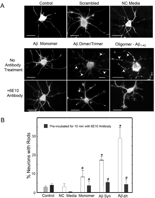
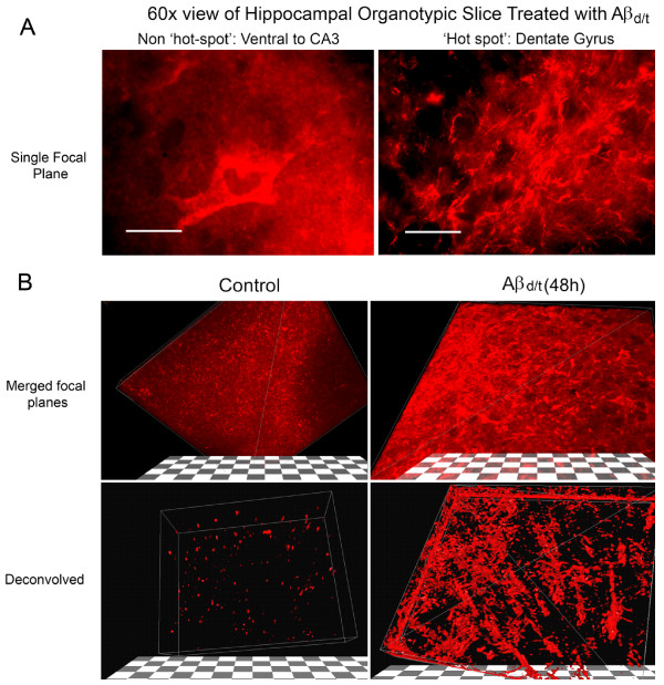
Background: Previously we reported 1 μM synthetic human amyloid beta1-42 oligomers induced cofilin dephosphorylation (activation) and formation of cofilin-actin rods within rat hippocampal neurons primarily localized to the dentate gyrus.

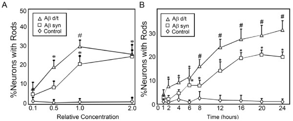
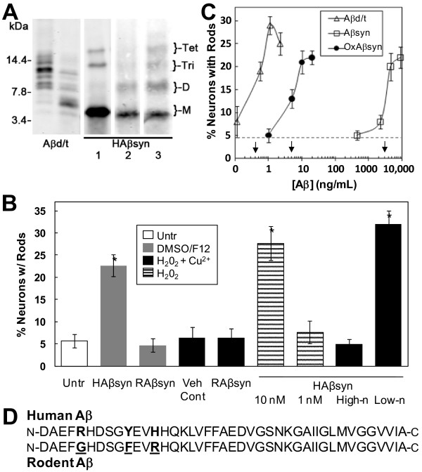
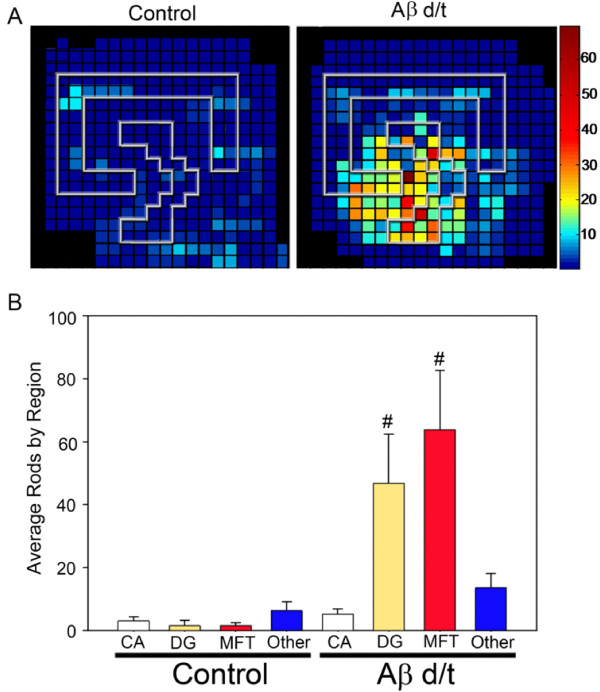
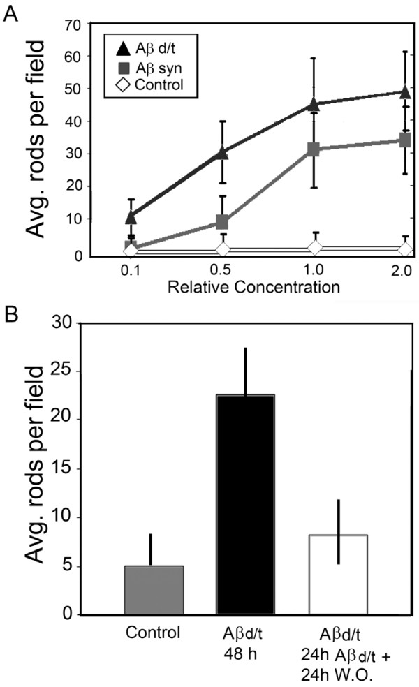
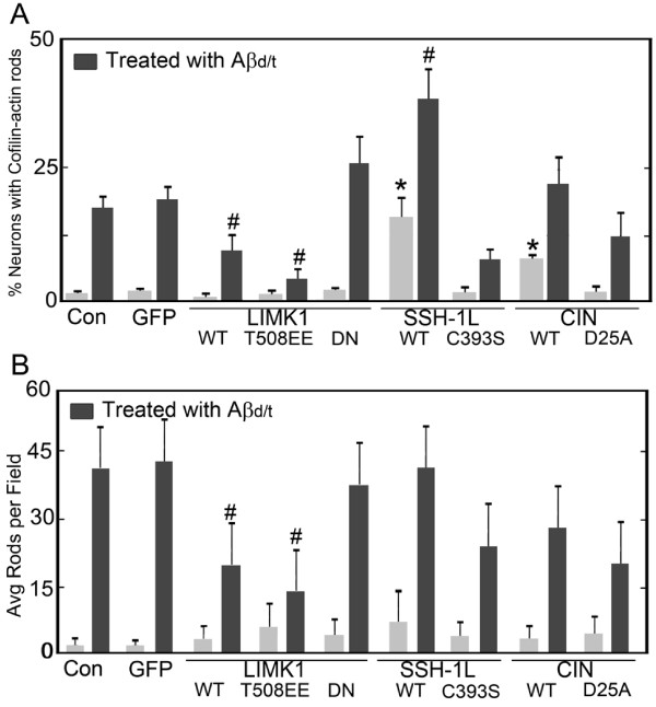
Results: Here we demonstrate that a gel filtration fraction of 7PA2 cell-secreted SDS-stable human Aβ dimers and trimers (Aβd/t) induces maximal neuronal rod response at ~250 pM. This is 4,000-fold more active than traditionally prepared human Aβ oligomers, which contain SDS-stable trimers and tetramers, but are devoid of dimers. When incubated under tyrosine oxidizing conditions, synthetic human but not rodent Aβ1-42, the latter lacking tyrosine, acquires a marked increase (620 fold for EC50) in rod-inducing activity. Gel filtration of this preparation yielded two fractions containing SDS-stable dimers, trimers and tetramers. One, eluting at a similar volume to 7PA2 Aβd/t, had maximum activity at ~5 nM, whereas the other, eluting at the void volume (high-n state), lacked rod inducing activity at the same concentration. Fractions from 7PA2 medium containing Aβ monomers are not active, suggesting oxidized SDS-stable Aβ1-42 dimers in a low-n state are the most active rod-inducing species. Aβd/t-induced rods are predominantly localized to the dentate gyrus and mossy fiber tract, reach significance over controls within 2 h of treatment, and are reversible, disappearing by 24 h after Aβd/t washout. Overexpression of cofilin phosphatases increase rod formation when expressed alone and exacerbate rod formation when coupled with Aβd/t, whereas overexpression of a cofilin kinase inhibits Aβd/t-induced rod formation.

Conclusions: Together these data support a mechanism by which Aβd/t alters the actin cytoskeleton via effects on cofilin in neurons critical to learning and memory.


Figures

Figure 1
Figure 1
Aβd/t fraction from 7PA2 cells, but not monomer, induces rods in dissociated hippocampal neurons. Analysis by fluorescence microscopy of dissociated neurons treated with vehicle (control), scrambled Aβ peptide (1 μM), or non-conditioned (NC media, d/t equivalent fraction), as well as with the monomer and d/t fractions from 7PA2 cell culture medium and 1 μM synthetic Aβ oligomers (Aβsyn). (A) Cofilin immunostained fluorescence images of hippocampal neurons showing representative responses with cofilin-actin rods (arrowheads) formed 24 h after treatment with Aβd/t and Aβsyn oligomers. Pretreatment of the Aβ-fractions with an antibody (6E10) to Aβ eliminates their rod-inducing effects. Bars = 10 μm. (B) Quantification of the rod forming response showing the neutralizing effects of the 6E10 antibody and the non-significant changes in rod formation by Aβ monomer. (* = p = 0.01 compared to control; # not significantly different from control).
Figure 2
Figure 2
Percent of neurons in dissociated hippocampal cultures containing rods as a function of Aβ form, concentration and time of treatment. (A) Dose-response curves for Aβsyn oligomers and Aβd/t versus control. The concentrations are expressed in terms of the 7PA2 CHO cell secreted concentration of Aβd/t (1X = 250 pM), which was used at 0.1, 0.5 and 2X this value. For the Aβsyn oligomers, the 1X value equals 1.0 μM. (B) Following treatment with 1X amounts of Aβd/t or Aβsyn oligomers, neurons were fixed at the times shown and the percent of neurons with rods was quantified. By 2 h the percent of neurons forming rods in response to Aβd/t was significant (*) over controls (p = 0.05). The Aβsyn-treated neurons required 6 h to reach significance over controls. Significance (# = p = 0.05) in the differences between the two Aβ species occurred at 8 h.
Figure 3
Figure 3
Oxidized cross-linking of synthetic Aβ1-42 generates a dimer that potently induces rods. (A) Western blot showing SDS-stable species of Aβ in different preparations. Two gel filtration fractions from medium of 7PA2 cells are combined to give Aβd/t. Last three lanes are synthetic human Aβ1-42. Lane 1: traditional oligomers prepared in DMSO/F12 [44,45]; Lane 2: peptide incubated (37°C, 3 d) in PBS containing 250 μM H2O2; Lane 3: peptide incubated (5 d) in PBS containing 250 μM peroxide plus 25 μM CuCl2. Dimer is absent in traditional oligomer preparations but forms in peroxide alone. Dimer, trimer and tetramer are generated with Cu2+/peroxide. (B) Comparison of rod-inducing activity between untreated (Untr) dissociated neurons and neurons treated with: Cu2+/peroxide (veh cont), 1 μM traditional synthetic human Aβ oligomers (HAβsyn), 1 μM synthetic rodent Aβ (RAβsyn) treated identically to HAβsyn, 1 μM Cu2+/peroxide-treated RAβsyn, 10 nM (45 ng/mL) and 1 nM peroxide-oxidized HAβsyn, and 5 nM each of high-n and low-n oligomers-containing fractions of oxidized HAβsyn (see Additional file 3). (C) The rod-inducing activity of different Aβ preparations. The effective concentration for a 50% maximal response (EC50; arrows) was calculated from dose-response curves for traditional Aβ oligomers (Aβsyn; ED50 = 3100 ng/mL), Cu2+/peroxide oxidized synthetic Aβ (OxAβsyn; ED50 = 5 ng/mL) and Aβd/t (ED50 = 0.4 ng/mL). Dashed line is control (untreated). Bars are standard deviations. (D) Human and rodent Aβ1-42 sequences differ in three residues (bold), but only tyrosine at position 10 is likely to generate the peroxide-induced SDS-stable species.
Figure 4
Figure 4
Numbers of rods induced by Aβd/t are highest in the dentate gyrus and mossy fiber tract. (A) Organotypic hippocampal slices were cultured for at least 8-10 days and were left untreated or treated with 1X Aβd/t, the same amount of the equivalent NC medium fraction, or scrambled Aβ peptide. After 48 h, slices were fixed and immunostained for cofilin and DNA (DAPI), and rods were quantified by counting with a 60x objective. Rod mapping from multiple slices onto a matrix grid of the hippocampus was performed as previously described using fiduciary markers from the stained nuclei layers to align hippocampal regions [29]. There were no differences detected in rod numbers or distribution between the untreated slices and those treated with NC medium or scrambled peptide and these were all combined to give the control panel. Rods induced by the Aβd/t were heavily concentrated over the dentate gyrus and mossy fiber tract (n = 18 for control slices and n = 12 for Aβd/t treated). (B) Rod quantification averaged per field over different regions of the slices. Each field acquired with the 60x objective has about 6-7 matrix grid squares. The only regions of significance (# = p = 0.05) for the rod numbers are in the dentate gyrus and mossy fiber tract.
Figure 5
Figure 5
Dose-response curve for rod formation in organotypic hippocampal slices and reversibility of Aβd/t-induced rods. (A) The same concentrations of Aβd/t and synthetic oligomer used in Figure 2A were applied to organotypic hippocampal slices. After 48 h, slices were fixed and rods immunostained and quantified per field averaged across the entire slice. The curves obtained are very similar to those in dissociated neurons but the measured parameters are different (average rods per field measured here vs. percent neurons with rods in Figure 2A). (B) Rods formed in response to Aβd/t in hippocampal slices reached their maximum value by 24 h (see Figure 2B). To determine rod reversibility, some of the slices were washed free of the Aβd/t and allowed to incubate another 24 h, whereas others had the Aβd/t present continuously. Controls were treated with the NC medium for 48 h or were left untreated (no difference). As previously shown for the rods induced by synthetic Aβ oligomers [29], the Aβd/t-induced rods are also reversible.
Figure 6
Figure 6
Three-dimensional reconstruction of cofilin-stained rods in deconvolved confocal image stacks from organotypic slices. Treatment of organotypic slices with Aβd/t results in a profound increase in cofilin-immunostained rods in the dentate gyrus/mossy fiber tract (DG/MFT) and a global change in cofilin distribution in cells in this region. (A) Single focal plane of non-rod-forming region near the CA3 compared to a rod hot spot in the dentate gyrus. Rods are evident in this single plane. (B) Three dimensional stack of planes from a cofilin stained control and Aβd/t-treated slice. Deconvolution of the confocal image stacks and thresholding the image by removing the lowest 20% of signal (lower panels) provides striking evidence of rod formation in this region. Hundreds of rods can be observed, which contain virtually all of the remaining immunostained cofilin.
Figure 7
Figure 7
Upstream regulators of cofilin phosphorylation impact the ability of Aβd/t to induce rods. Rod formation was quantified in (A) dissociated hippocampal neuronal cultures or (B) organotypic hippocampal slices that were uninfected (Con), infected with control adenovirus expressing GFP (GFP) or with adenoviruses expressing various upstream regulators of cofilin phosphorylation. All viruses co-expressed a fluorescent protein marker and only neurons expressing the marker were scored in the dissociated cultures. In slices, infection rates were approximately 70% (see Additional file 5) and rods per field were quantified (it was not possible in slices to count rods only within infected neurons). Neurons or slices were infected 24 h prior to treatment with Aβd/t (1X) and were fixed and analyzed for rod formation 48 h after Aβd/t addition. Treatments that enhance cofilin dephosphorylation (the active phosphatases SSH-1L WT and CIN WT) in the dissociated cultures enhance rod formation with or without Aβd/t treatment (* = significant difference from untreated or GFP controls at p = 0.05; # significantly different from Aβd/t treated controls, p = 0.05). Treatments that inhibit cofilin dephosphorylation (LIMK1 WT and the active LIMK1T508EE), inhibit rod formation in response to Aβd/t in both dissociated neuronal cultures and in slices (n = 3).

Similar articles

Cited by

References

    1. Glenner GG, Wong CW. Alzheimer's disease: initial report of the purification and characterization of a novel cerebrovascular amyloid protein. Biochem Biophys Res Commun. 1984;120:885–890. doi: 10.1016/S0006-291X(84)80190-4. - DOI - PubMed
    1. Hardy J, Selkoe DJ. The amyloid hypothesis of Alzheimer's disease: progress and problems on the road to therapeutics. Science. 2002;297:353–356. doi: 10.1126/science.1072994. - DOI - PubMed
    1. Mattson MP. Pathways towards and away from Alzheimer's disease. Nature. 2004;430:631–639. doi: 10.1038/nature02621. - DOI - PMC - PubMed
    1. Price DL, Sisodia SS, Gandy SE. Amyloid beta amyloidosis in Alzheimer's disease. Curr Opin Neurol. 1995;8:268–274. doi: 10.1097/00019052-199508000-00004. - DOI - PubMed
    1. Sisodia SS, Price DL. Role of the beta-amyloid protein in Alzheimer's disease. FASEB J. 1995;9:366–370. - PubMed

LinkOut - more resources