

Development and evaluation of a virtual microscopy application for automated assessment of Ki-67 expression in breast cancer
- PMID: 21262004
- PMCID: PMC3040126
- DOI: 10.1186/1472-6890-11-3
Development and evaluation of a virtual microscopy application for automated assessment of Ki-67 expression in breast cancer
Abstract
Background: The aim of the study was to develop a virtual microscopy enabled method for assessment of Ki-67 expression and to study the prognostic value of the automated analysis in a comprehensive series of patients with breast cancer.
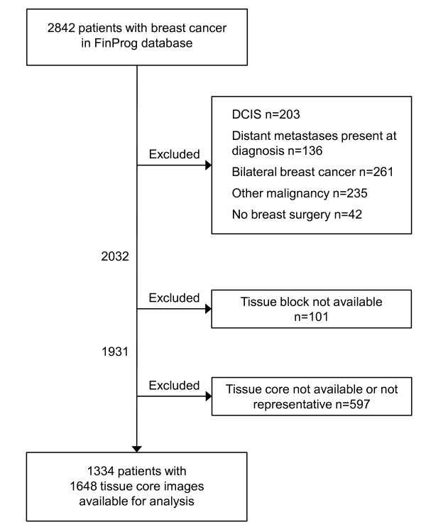
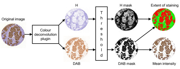
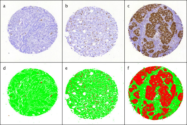
Methods: Using a previously reported virtual microscopy platform and an open source image processing tool, ImageJ, a method for assessment of immunohistochemically (IHC) stained area and intensity was created. A tissue microarray (TMA) series of breast cancer specimens from 1931 patients was immunostained for Ki-67, digitized with a whole slide scanner and uploaded to an image web server. The extent of Ki-67 staining in the tumour specimens was assessed both visually and with the image analysis algorithm. The prognostic value of the computer vision assessment of Ki-67 was evaluated by comparison of distant disease-free survival in patients with low, moderate or high expression of the protein.
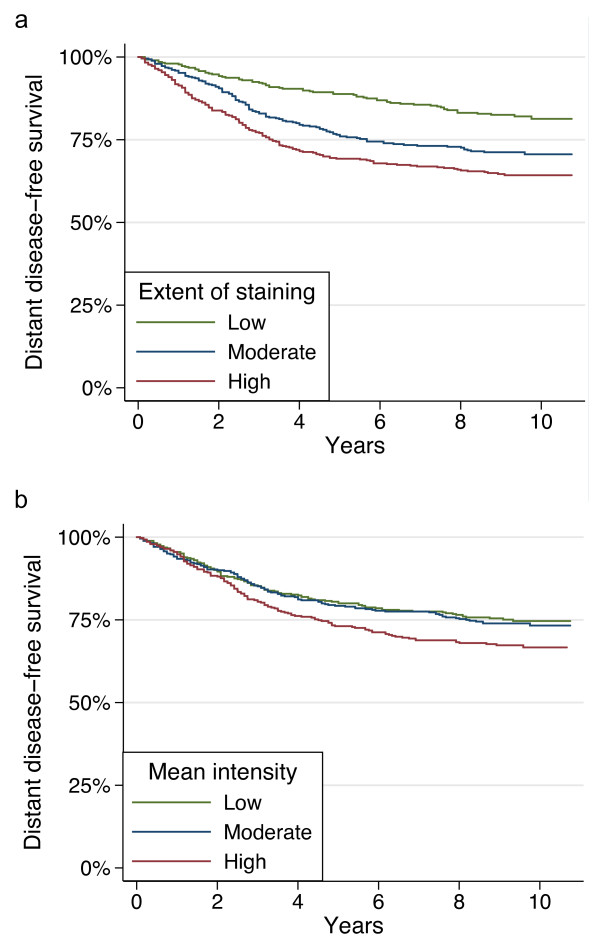
Results: 1648 evaluable image files from 1334 patients were analysed in less than two hours. Visual and automated Ki-67 extent of staining assessments showed a percentage agreement of 87% and weighted kappa value of 0.57. The hazard ratio for distant recurrence for patients with a computer determined moderate Ki-67 extent of staining was 1.77 (95% CI 1.31-2.37) and for high extent 2.34 (95% CI 1.76-3.10), compared to patients with a low extent. In multivariate survival analyses, automated assessment of Ki-67 extent of staining was retained as a significant prognostic factor.
Conclusions: Running high-throughput automated IHC algorithms on a virtual microscopy platform is feasible. Comparison of visual and automated assessments of Ki-67 expression shows moderate agreement. In multivariate survival analysis, the automated assessment of Ki-67 extent of staining is a significant and independent predictor of outcome in breast cancer.
Figures
References
LinkOut - more resources
Full Text Sources
