. 2011 Apr;140(4):1219-1229.e1-2.
doi: 10.1053/j.gastro.2011.01.033. Epub 2011 Jan 21.

GATA factors regulate proliferation, differentiation, and gene expression in small intestine of mature mice

Affiliations

GATA factors regulate proliferation, differentiation, and gene expression in small intestine of mature mice

Eva Beuling et al. Gastroenterology. 2011 Apr.

Abstract

Background & aims: GATA transcription factors regulate proliferation, differentiation, and gene expression in multiple organs. GATA4 is expressed in the proximal 85% of the small intestine and regulates the jejunal-ileal gradient in absorptive enterocyte gene expression. GATA6 is co-expressed with GATA4 but also is expressed in the ileum; its function in the mature small intestine is unknown.

Methods: We investigated the function of GATA6 in small intestine using adult mice with conditional, inducible deletion of Gata6, or Gata6 and Gata4, specifically in the intestine.

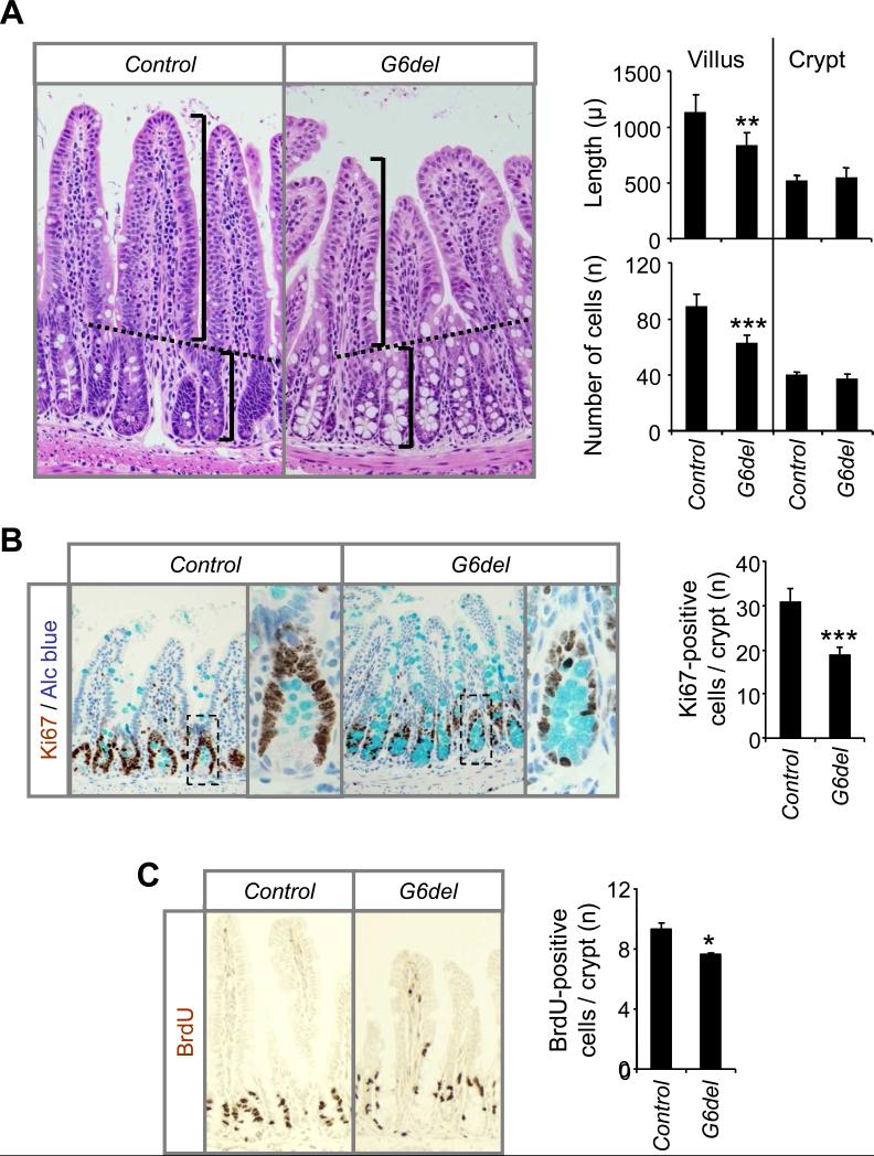
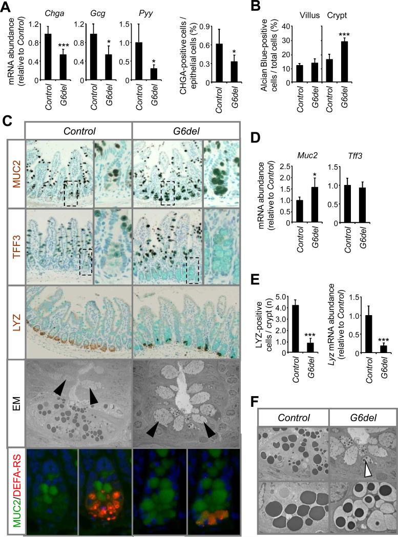
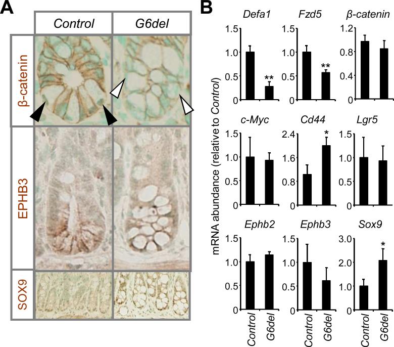
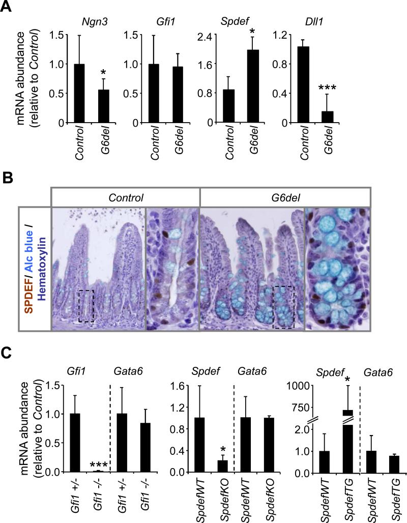
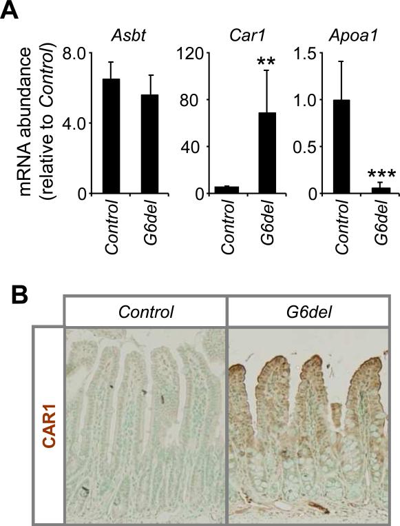
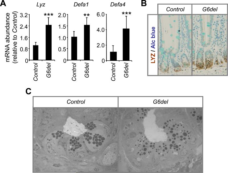
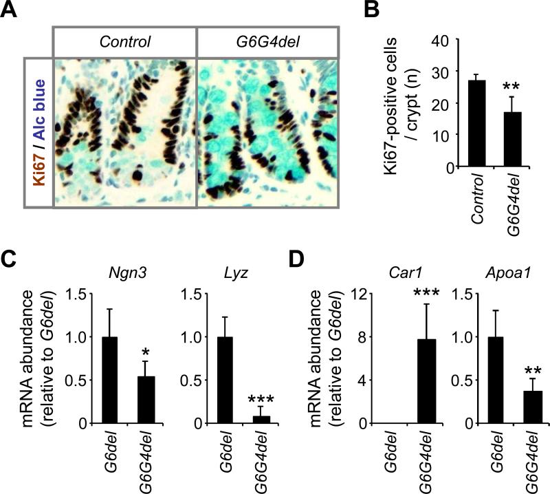
Results: In ileum, deletion of Gata6 caused a decrease in crypt cell proliferation and numbers of enteroendocrine and Paneth cells, an increase in numbers of goblet-like cells in crypts, and altered expression of genes specific to absorptive enterocytes. In contrast to ileum, deletion of Gata6 caused an increase in numbers of Paneth cells in jejunum and ileum. Deletion of Gata6 and Gata4 resulted in a jejunal and duodenal phenotype that was nearly identical to that in the ileum after deletion of Gata6 alone, revealing common functions for GATA6 and GATA4.

Conclusions: GATA transcription factors are required for crypt cell proliferation, secretory cell differentiation, and absorptive enterocyte gene expression in the small intestinal epithelium.


Figures

Figure 1
Figure 1
Intestinal Gata6 deletion results in a reduction in proliferation in the mature ileum. (A) From hematoxylin and eosin stained slides, crypt-villus junctions were established (dotted line) and villus height and crypt depth (brackets), as well as villus and crypt cell number, were determined (left panel) as indicated in Materials and Methods. Villus height and cell number were decreased in G6del as compared to Control ileum (right panel). (**P<.01, ***P<.001, as compared to Control). (B) Immunostaining for Ki67 (left panel) reveals that the number of positive cells was decreased after Gata6 deletion (right panel). (***P<.001, as compared to Control). (C) Immunohistochemistry for bromodeoxyuridine (BrdU) (left panel) demonstrates a decrease in the number of positive cells after Gata6 deletion (left panel). (*P<.05, as compared to Control, n=2 in each group).
Figure 2
Figure 2
Intestinal Gata6 deletion results in an increase in goblet-like cells in crypts, and a decrease in Paneth and enteroendocrine cells. (A) Quantitative RT-PCR shows a decrease in enteroendocrine marker transcripts; number of CHGA-positive cells are decreased in G6del ileum. (B) Number alcian blue-positive cells is increased in crypts. (C) Immunostaining for MUC2, TFF3 and LYZ shows an increase and MUC2 and decrease in LYZ in crypts; electron microscopy shows an increase in goblet-like cells in crypts; co-immunofluorescence shows co-localization of DEFA-RS and MUC2 in crypts of G6del mice. (D) Quantitative RT-PCR of Muc2 and Tff3 shows an increase in Muc2 mRNA abundance. (E) Number of LYZ-positive cells in crypts and Lyz mRNA abundance are both decreased in G6del ileum. (F) Electron microscopy shows the presence of cells with mixed goblet/Paneth granules in G6del ileum. *P<.05, **P<.01, ***P<.001, as compared to Control.
Figure 3
Figure 3
Intestinal Gata6 deletion results in changes in the expression of crypt Wnt targets in ileum. (A) Immunostaining reveals a decrease in nulcear β-catenin, expression of EPHB3, and an increase in intensity of SOX9 in crypts of G6del ileum. (B) Quantitative RT-PCR shows changes in the mRNA abundance of specific proteins involved in intestinal Wnt signaling. (*P<.05, **P<.01, as compared to Control).
Figure 4
Figure 4
Intestinal Gata6 deletion results in alterations in Notch signaling targets. (A) Quantitative RT-PCR shows changes in the mRNA abundance of specific proteins involved in intestinal Notch signaling. (B) Immunostaining reveals an increase in SPDEF in crypts of G6del ileum. (C) Quantitative RT-PCR shows no change in the mRNA abundance of Gata6 in Gfi1 knockout (Gfi1-/-), Spdef knockout (SpdefKO) and Spdef over-expressing (SpdefTG) mice. *P<.05, **P<.01, ***P<.001, as compared to Control.
Figure 5
Figure 5
Intestinal Gata6 deletion results in alterations in the expression of specific absorptive enterocyte genes. (A) Quantitative RT-PCR shows no change in Asbt, an increase in Car1, and a decrease in Apoa1 mRNA abundances in G6del. **P<.01, ***P<.001, as compared to Control. (B) Immunostaining shows an increase in CAR1 in absorptive enterocytes of G6del ileum.
Figure 6
Figure 6
Intestinal Gata6 deletion results in an increase in Paneth cells in the jejunum. (A) Quantitative RT-PCR shows increases in Lyz, Defa1 and Defa4 mRNA abundance after Gata6 deletion. **P<.01, ***P<.001, as compared to Control. (B) Immunohistochemistry revealed an increased intensity and a more widespread pattern of LYZ staining in jejunal crypts after Gata6 deletion. (C) Electron microscopy revealed more Paneth cells and Paneth granules that were more variable in size and more widely dispersed in G6del ileum.
Figure 7
Figure 7
GATA4 is redundant for most GATA6 functions in the mature jejunum. (A) Sections stained for Ki67 and alcian blue revealed (B) a decrease in the number of Ki67-positive cells per crypt in G6G4del as compared to Control jejunum. (C) Quantitative RT-PCR shows decreases in Ngn3 and Lyz mRNA abundances in G6G4del jejunum. (D) Quantitative RT-PCR shows an increase in Car1 and a decrease in Apoa1 mRNA abundance in G6G4del jejunum. *P<.05, **P<.01, ***P<.001, as compared to G6del jejunum.

Similar articles

Cited by

References

    1. Stappenbeck TS, Wong MH, Saam JR, et al. Notes from some crypt watchers: regulation of renewal in the mouse intestinal epithelium. Curr Opin Cell Biol. 1998;10:702–9. - PubMed
    1. Ireland H, Kemp R, Houghton C, et al. Inducible Cre-mediated control of gene expression in the murine gastrointestinal tract: effect of loss of beta-catenin. Gastroenterology. 2004;126:1236–46. - PubMed
    1. Korinek V, Barker N, Moerer P, et al. Depletion of epithelial stem-cell compartments in the small intestine of mice lacking Tcf-4. Nat Genet. 1998;19:379–83. - PubMed
    1. Kuhnert F, Davis CR, Wang HT, et al. Essential requirement for Wnt signaling in proliferation of adult small intestine and colon revealed by adenoviral expression of Dickkopf-1. Proc Natl Acad Sci U S A. 2004;101:266–71. - PMC - PubMed
    1. Pinto D, Gregorieff A, Begthel H, et al. Canonical Wnt signals are essential for homeostasis of the intestinal epithelium. Genes Dev. 2003;17:1709–13. - PMC - PubMed

Publication types

MeSH terms