. 2011 Mar 18;407(1):149-70.
doi: 10.1016/j.jmb.2011.01.034. Epub 2011 Jan 22.

The DNA-binding domain of human PARP-1 interacts with DNA single-strand breaks as a monomer through its second zinc finger

Affiliations

The DNA-binding domain of human PARP-1 interacts with DNA single-strand breaks as a monomer through its second zinc finger

Sebastian Eustermann et al. J Mol Biol. .

Erratum in

  • J Mol Biol. 2012 Jun 8;419(3-4):275-6

Abstract

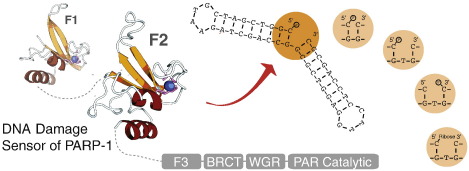
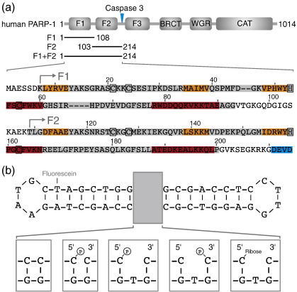
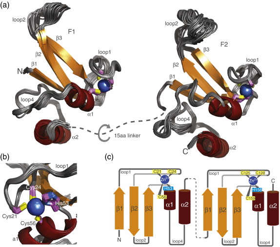
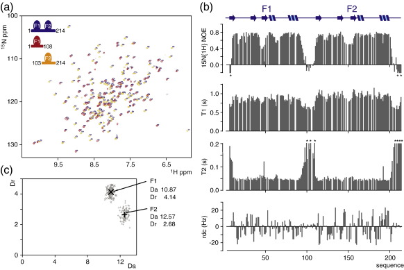
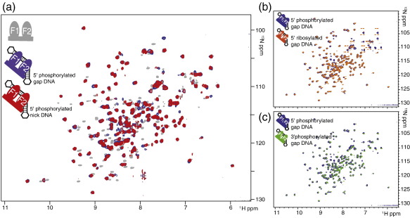
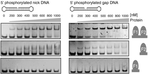
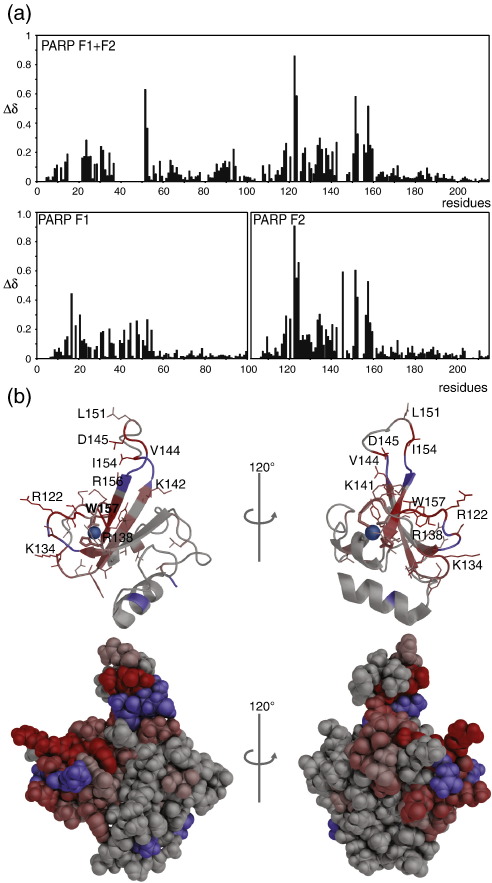
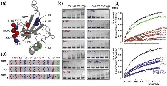
Poly(ADP-ribose)polymerase-1 (PARP-1) is a highly abundant chromatin-associated enzyme present in all higher eukaryotic cell nuclei, where it plays key roles in the maintenance of genomic integrity, chromatin remodeling and transcriptional control. It binds to DNA single- and double-strand breaks through an N-terminal region containing two zinc fingers, F1 and F2, following which its C-terminal catalytic domain becomes activated via an unknown mechanism, causing formation and addition of polyadenosine-ribose (PAR) to acceptor proteins including PARP-1 itself. Here, we report a biophysical and structural characterization of the F1 and F2 fingers of human PARP-1, both as independent fragments and in the context of the 24-kDa DNA-binding domain (F1+F2). We show that the fingers are structurally independent in the absence of DNA and share a highly similar structural fold and dynamics. The F1+F2 fragment recognizes DNA single-strand breaks as a monomer and in a single orientation. Using a combination of NMR spectroscopy and other biophysical techniques, we show that recognition is primarily achieved by F2, which binds the DNA in an essentially identical manner whether present in isolation or in the two-finger fragment. F2 interacts much more strongly with nicked or gapped DNA ligands than does F1, and we present a mutational study that suggests origins of this difference. Our data suggest that different DNA lesions are recognized by the DNA-binding domain of PARP-1 in a highly similar conformation, helping to rationalize how the full-length protein participates in multiple steps of DNA single-strand breakage and base excision repair.


Figures

None
Graphical abstract
Fig. 1
Fig. 1
Protein and DNA constructs used in this study. (a) During apoptosis, PARP-1 is cleaved by caspase-3 into a 24-kDa fragment that contains the two N-terminal zinc fingers (F1 and F2) and an 89-kDa fragment composed of the third zinc finger (F3), the BRCT domain, the WGR domain and the C-terminal catalytic domain (the caspase-3 cleavage site is marked with a blue arrow). The expansion below shows the sequence of human PARP-1 residues 1–214, highlighting the fragments used in this study: F1 (residues 1–108), F2 (residues 103–214) and F1 + F2 (residues 1–214). Secondary structural elements are colored (α-helices, red; β-strands, orange), the caspase-3 recognition site is shown in blue and zinc-coordinating residues are indicated by black boxes. (b) Synthetic DNA dumbbell ligands used in this study. The same dumbbell scaffold was used to harbour different types of DNA strand breaks: a 5′-phosphorylated nick, a 5′-phosphorylated single nucleotide gap, a 3′-phosphorylated single nucleotide gap and a 5′-ribosylated single nucleotide gap (which results from strand incision of an abasic site during BER). Ligation of the nicked DNA dumbbell ligand produced a circular DNA without a strand break (left-handmost box).
Fig. 2
Fig. 2
Solution structures of the N-terminal zinc fingers F1 and F2 of human PARP-1. (a) Ensemble views of the 30 accepted structures of F1 (backbone rmsd 0.39 Å) and F2 (backbone rmsd 0.37 Å) shown in a cartoon representation, colored as for Fig. 1. Zinc ions are shown as blue spheres and zinc coordinating residues in stick representation with carbon atoms in magenta. The two zinc fingers are connected by a linker of 15 residues (shown as a dotted line) that is flexible, as judged by NMR measurements of PARP-1 F1 + F2. (b) Close-up of the zinc coordination of PARP-1 F1 [rotated about the vertical by 90° relative to the view in (a)]. The absolute chirality of the zinc binding configuration is S, as defined by Berg. (c) Schematic showing the zinc binding and secondary-structure topology of the two N-terminal PARP-1 zinc finger domains F1 and F2, colored as for Fig. 1.
Fig. 3
Fig. 3
Fingers F1 and F2 of human PARP-1 are structurally independent. (a) Overlay of [15N–1H] HSQC spectra of PARP-1 fragments F1 (red peaks), F2 (yellow peaks) and F1 + F2 (blue peaks). To help visualize the relationships, we deliberately introduced a small systematic chemical shift offset for the spectrum of PARP-1 F1 + F2. (b) Internal motions of backbone amides of PARP-1 F1 + F2 were assessed by NMR spectroscopy. Steady-state 15N{1H} NOE values, 15N T1 and 15N T2 relaxation times and amide group RDC values are each plotted as a function of sequence. The relaxation data (upper three panels) demonstrate that the linker region between the two zinc fingers F1 and F2 (residues 94–108) is flexible, having internal motions that are faster than overall molecular tumbling; in addition, significant motions were detected for the protein termini and for some loop regions of each zinc finger (described in the main text). The RDC data (lowest panel) were recorded from a sample of PARP-1 F1 + F2 weakly aligned by pf1 phage, using [15N–1H] HSQC IPAP (in-phase antiphase) spectra. Larger values are only seen for the ordered regions. (c) Using the RDCs measured for PARP-1 F1 + F2 and the solution structure of the separated F1 and F2, we determined the axial (Da) and rhombic (Dr) components of the alignment tensor of each finger in the context of a two-finger construct. Associated uncertainties were estimated using the program MODULE 2.0, setting the error of measurement for experimentally determined RDCs to 2 Hz (see Supplementary Material and Methods). The unrelated alignment tensors of F1 and F2 provide additional evidence for the lack of any rigid orientation between the two zinc fingers.
Fig. 4
Fig. 4
Biophysical characterization of PARP-1 binding to DNA single-strand breaks. (a) Sedimentation velocity experiments were carried out using 0.5 μM nicked dumbbell DNA alone or in the presence of a fivefold excess of PARP-1 F1 + F2. As a control, the latter experiment was repeated using a ligated version of the DNA dumbbell. Raw data were fitted as described in Supplementary Fig. 2, and diffusion-deconvoluted sedimentation coefficient distributions c(s) are plotted for each experiment. The c(s) distribution of the nicked dumbbell DNA + PARP-1 F1 + F2 sample corresponds to that expected for formation of a homogenous 1:1 protein–DNA complex (fitted molecular mass 43.5 kDa; calculated molecular mass 38 kDa). (b) Stochiometric fluorescence anisotropy titrations of 44 nt 5′-phosphorylated fluorescently labeled DNA (1 μM) and PARP-1 F1 + F2 (0–6.25 μM) were carried out using a buffer that contained either 0 mM (blue circles) or 200 mM sodium chloride (red circles). Stochiometric points were deduced as indicated. (c–e) To determine single-strand nick-specific and nonspecific DNA binding affinities for PARP-1 F1 + F2, we carried out fluorescence anisotropy titrations at a DNA concentration of 10 nM. The following DNA ligands were used: (c) 44 nt 5′-phosphorylated fluorescently labeled nicked DNA dumbbell; (d) 44 nt 5′-phosphorylated fluorescently labeled nicked DNA dumbbell with symmetric stem structures; (e) same ligand as in (d) but ligated to form a circular DNA (see Supplementary Table 2 for DNA sequences). Experiments were carried out at increasing ionic strengths (0, 25, 50, 75, 100, 150 and 200 mM sodium chloride), binding data are colored stepwise from blue to red and data fits are shown as continuous lines. (f) Fitted KD dissociation constants for single-strand nick-specific (KD1) and nonspecific binding (KD and KD2) are plotted as a function of ionic strength (numerical values for the fitted dissociation constants are given in Supplementary Table 1). Data were analyzed using either a one-binding-site model or a two-binding-site model as appropriate. Binding to symmetrically ligated DNA was not detected at 150 and 200 mM NaCl (e) and was therefore only analyzed at sodium chloride concentrations up to 100 mM.
Fig. 5
Fig. 5
Interaction of PARP-1 F1 + F2 with different types of single-strand breaks. (a) Overlay of [15N–1H] HSQC spectra of PARP-1 fragment F1 + F2 either in the free state (gray peaks), bound to a 45-nt 5′-phosphorylated gapped DNA dumbbell (blue peaks) or to a 44-nt 5′-phosphorylated nicked DNA dumbbell (red peaks). (b and c) Blue peaks belong to the same spectrum as in (a) overlaid with [15N–1H] TROSY spectra of PARP-1 fragment F1 + F2 (70% deuterated) bound to either a 45-nt 5′ ribosylated gapped DNA dumbbell (orange peaks) or a 45-nt 3′-phosphorylated gapped DNA dumbbell (green peaks). Closely corresponding peak positions of the DNA-bound proteins strongly indicate that PARP-1 F1 + F2 recognizes all these three types of DNA single-strand breaks in a very similar manner.
Fig. 6
Fig. 6
Gel electromobility shift assays. Electromobility gel shift assays of 44-nt 5′-phosphorylated nicked DNA dumbbell (left panel) and 45-nt 5′-phosphorylated gapped DNA dumbbell (right panel) binding to PARP-1 fragments. F1 + F2 binds the most strongly, while F2 binds slightly less strongly, and binding of F1 is not detected in these experiments. Binding to the gapped ligand is consistently slightly stronger than to the nicked ligand. EMSA experiments were performed using 400 nM DNA and increasing protein concentrations (see labeling) of PARP-1 F1 + F2, F2 and F1 (for each binding reaction a volume of 10 μl was loaded). The gel shift experiments allowed a comparison between the different complexes, but binding constants are probably underestimated in comparison to those seen in fluorescence anisotropy measurements due to the nonequilibrium nature of the experiment.
Fig. 7
Fig. 7
Comparison of F2 and F1 + F2 in interaction with DNA single-strand breaks. Overlay of [15N–1H] HSQC spectra of PARP-1 fragment F1 + F2 and F2 either in the free state (F2, gray peaks; F1 + F2, black peaks) or in the DNA-bound state (F2, orange peaks; F1 + F2, blue peaks). Two expansions are shown in (a) and (b). The two 1:1 protein–DNA complexes (100 μM) were each reconstituted under the same experimental conditions using the 44-nt 5′-phosphorylated DNA dumbbell with a single-stranded nick. DNA binding caused essentially identical chemical shift perturbations for F2 alone (red arrows) as it did for F2 in the context of a two-zinc-finger construct (F1 + F2; blue arrows), as exemplified in the expansions. These data demonstrate that PARP-1 F2 retains the same DNA binding configuration regardless of the presence or absence of F1.
Fig. 8
Fig. 8
Chemical shift perturbation analysis. (a) Histograms of backbone amide chemical shift perturbations for F1 + F2 (upper panel), F1 and F2 (both lower panels) upon binding to the 45-nt 5′-phosphorylated gapped DNA dumbbell. Perturbations are calculated as Δδ = √[(δ1H)2 + (δ15N ÷ 5)2] (b) Chemical shift perturbations from (a) mapped onto the solution structure of F2 in both cartoon and space-filling representations. Perturbations were divided into five categories according to the SD over all perturbations: unaffected (0–0.5 × SD), weak (0.5–1 × SD), medium (1–1.5 × SD), strong (1.5–2 × SD) and very strong (> 2 × SD) and colored accordingly from gray (unaffected) to red (very strong). On the cartoon view, side chains of perturbed residues are drawn as thin lines. Residues for which no chemical shift perturbation could be determined due to missing assignments are colored blue.
Fig. 9
Fig. 9
Mutational analysis of PARP-1 F1 and F2. Two groups of mutations were selected. The first group (shown in red) includes residues that are similarly conserved between PARP-1 F1, PARP-1 F2 and the closely related zinc finger of DL3; mutations of these residues abolishes DNA-binding in every case (except K134I where it is significantly reduced). The second group (shown in blue) includes residues that are similarly conserved in PARP-1 F2 and DL3, but different in PARP-1 F1; single mutations in this group reduce DNA-binding affinity, while selected double mutations abolish it. As a control (shown in green), we mutated Lys197, which is not part of the DNA binding surface (see Fig. 8). (a) Mutations shown on the solution structure of PARP-1 F2. (b) Sequence logos produced using the multiple alignment of PARP-1 F1, PARP-1 F2 and DL3 given in Ref. . Residues Arg122 and Ser120 (F2 numbering) are conserved in PARP-1 F1 and F2 but transposed in DL3; however, they could still play corresponding roles in all three fingers. (c and d) DNA binding of wild-type PARP-1 F2 and mutants was compared using electrophoretic mobility gel shifts (c) and fluorescence anisotropy titrations (d). The gel experiments used the 45-nt 5′-phosphorylated DNA dumbbell ligand, and the fluorescence anisotropy titrations used fluorescently labeled 44-nt 5′-phosphorylated DNA dumbbell ligand (25 nM) in a buffer containing 50 mM Tris (pH 7), 25 mM NaCl, 150 μM ZnSO4 and 4 mM DTT. Fluorescence anisotropy data were analyzed using a one-binding-site model and the resulting apparent dissociation constants are given in Supplementary Table 2.

References

    1. D'Amours D., Desnoyers S., D'Silva I., Poirier G.G. Poly(ADP-ribosyl)ation reactions in the regulation of nuclear functions. Biochem. J. 1999;342(Pt 2):249–268. - PMC - PubMed
    1. Ame J.C., Spenlehauer C., de Murcia G. The PARP superfamily. Bioessays. 2004;26:882–893. - PubMed
    1. Adamietz P. Poly(ADP-ribose) synthase is the major endogenous nonhistone acceptor for poly(ADP-ribose) in alkylated rat hepatoma cells. Eur. J. Biochem. 1987;169:365–372. - PubMed
    1. Poirier G.G., de Murcia G., Jongstra-Bilen J., Niedergang C., Mandel P. Poly(ADP-ribosyl)ation of polynucleosomes causes relaxation of chromatin structure. Proc. Natl Acad. Sci. USA. 1982;79:3423–3427. - PMC - PubMed
    1. Tanuma S., Johnson L.D., Johnson G.S. ADP-ribosylation of chromosomal proteins and mouse mammary tumor virus gene expression. Glucocorticoids rapidly decrease endogenous ADP-ribosylation of nonhistone high mobility group 14 and 17 proteins. J. Biol. Chem. 1983;258:15371–15375. - PubMed

Publication types