

Nucleation and growth of mineralized bone matrix on silk-hydroxyapatite composite scaffolds
- PMID: 21262535
- PMCID: PMC3042545
- DOI: 10.1016/j.biomaterials.2010.12.058
Nucleation and growth of mineralized bone matrix on silk-hydroxyapatite composite scaffolds
Abstract
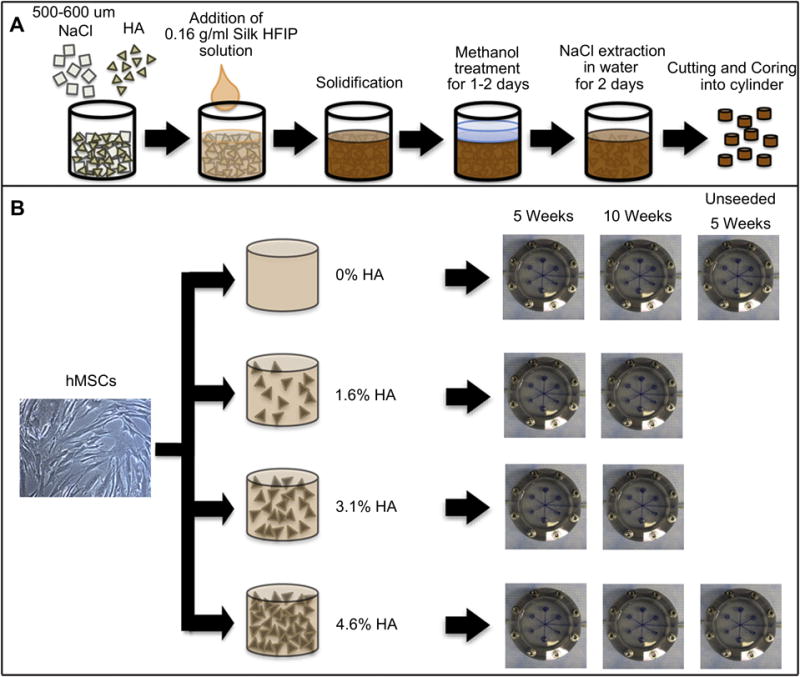
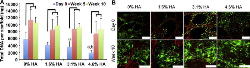
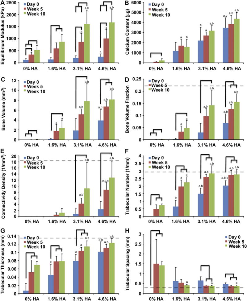
We describe a composite hydroxyapatite (HA)-silk fibroin scaffold designed to induce and support the formation of mineralized bone matrix by human mesenchymal stem cells (hMSCs) in the absence of osteogenic growth factors. Porous three-dimensional silk scaffolds were extensively used in our previous work for bone tissue engineering and showed excellent biodegradability and biocompatibility. However, silk is not an osteogenic material and has a compressive stiffness significantly lower than that of native bone. In the present study, we explored the incorporation of silk sponge matrices with HA (bone mineral) micro-particles to generate highly osteogenic composite scaffolds capable of inducing the in vitro formation of tissue-engineered bone. Different amounts of HA were embedded in silk sponges at volume fractions of 0%, 1.6%, 3.1% and 4.6% to enhance the osteoconductive activity and mechanical properties of the scaffolds. The cultivation of hMSCs in the silk/HA composite scaffolds under perfusion conditions resulted in the formation of bone-like structures and an increase in the equilibrium Young's modulus (up to 4-fold or 8-fold over 5 or 10 weeks of cultivation, respectively) in a manner that correlated with the initial HA content. The enhancement in mechanical properties was associated with the development of the structural connectivity of engineered bone matrix. Collectively, the data suggest two mechanisms by which the incorporated HA enhanced the formation of tissue engineered bone: through osteoconductivity of the material leading to increased bone matrix production, and by providing nucleation sites for new mineral resulting in the connectivity of trabecular-like architecture.
Copyright © 2011 Elsevier Ltd. All rights reserved.
Conflict of interest statement
Conflict of Interest: Authors declare no conflict of interest.
Figures


References
-
- Khan Y, Yaszemski MJ, Mikos AG, Laurencin CT. Tissue engineering of bone: material and matrix considerations. J Bone Joint Surg Am. 2008;90 1:36–42. - PubMed
-
- Griffith LG, Naughton G. Tissue engineering--current challenges and expanding opportunities. Science. 2002;295(5557):1009–14. - PubMed
-
- Langer R, Vacanti JP. Tissue engineering. Science. 1993;260(5110):920–6. - PubMed
-
- Kim HJ, Kim UJ, Vunjak-Novakovic G, Min BH, Kaplan DL. Influence of macroporous protein scaffolds on bone tissue engineering from bone marrow stem cells. Biomaterials. 2005;26(21):4442–52. - PubMed
-
- Martin I, Shastri VP, Padera RF, Yang J, Mackay AJ, Langer R, et al. Selective differentiation of mammalian bone marrow stromal cells cultured on three-dimensional polymer foams. J Biomed Mater Res. 2001;55(2):229–35. - PubMed
Publication types
MeSH terms
Substances
Grants and funding
LinkOut - more resources
Full Text Sources
Other Literature Sources
