. 2011 Apr;31(7):1551-64.
doi: 10.1128/MCB.00834-10. Epub 2011 Jan 24.

Tctex-1, a novel interaction partner of Rab3D, is required for osteoclastic bone resorption

Affiliations

Tctex-1, a novel interaction partner of Rab3D, is required for osteoclastic bone resorption

Nathan J Pavlos et al. Mol Cell Biol. 2011 Apr.

Abstract

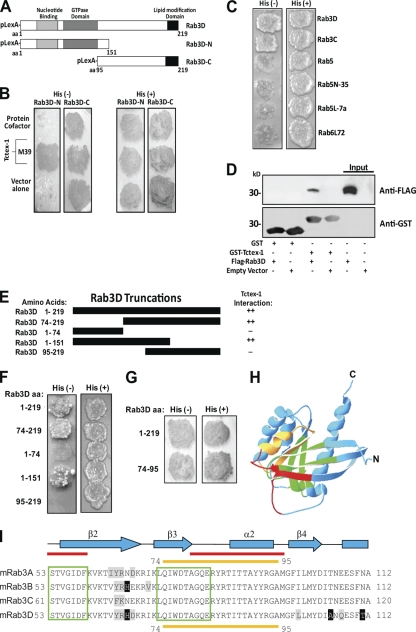
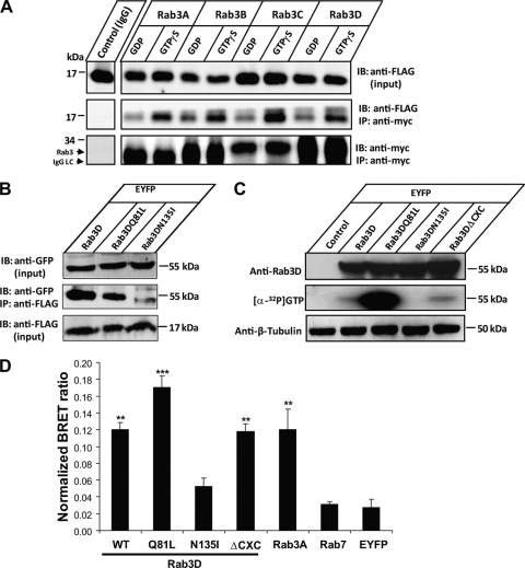
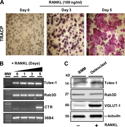
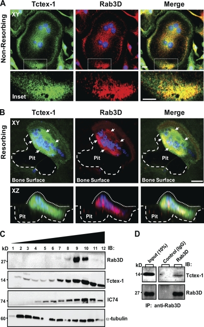
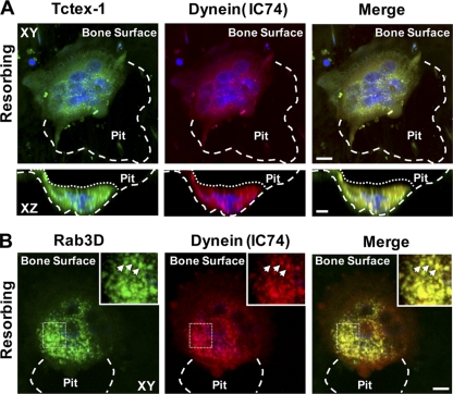
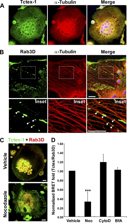
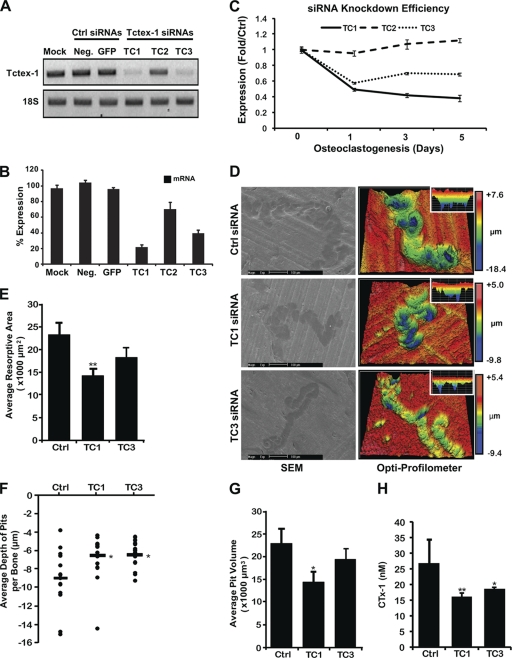
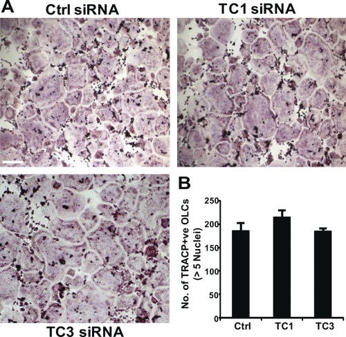
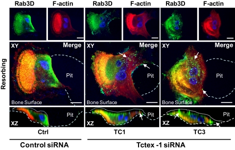
Vesicular transport along microtubules must be strictly regulated to sustain the unique structural and functional polarization of bone-resorbing osteoclasts. However, the molecular mechanisms bridging these vesicle-microtubule interactions remain largely obscure. Rab3D, a member of the Rab3 subfamily (Rab3A/B/C/D) of small exocytotic GTPases, represents a core component of the osteoclastic vesicle transport machinery. Here, we identify a new Rab3D-interacting partner, Tctex-1, a light chain of the cytoplasmic dynein microtubule motor complex, by a yeast two-hybrid screen. We demonstrate that Tctex-1 binds specifically to Rab3D in a GTP-dependent manner and co-occupies Rab3D-bearing vesicles in bone-resorbing osteoclasts. Furthermore, we provide evidence that Tctex-1 and Rab3D intimately associate with the dynein motor complex and microtubules in osteoclasts. Finally, targeted disruption of Tctex-1 by RNA interference significantly impairs bone resorption capacity and mislocalizes Rab3D vesicles in osteoclasts, attesting to the notion that components of the Rab3D-trafficking pathway contribute to the maintenance of osteoclastic resorptive function.


Figures

Fig. 1.
Fig. 1.
Identification of Tctex-1 as Rab3D binding protein. (A) Schematic representation of Rab3D N-terminal (aa 1 to 151) and C-terminal (aa 95 to 219) pBTM116 bait constructs used in the Y-2H screen. (B) Rab3D-N (aa 1 to 151) and Rab3D-C (aa 95 to 219) baits were cotransformed with VP16-protein cofactor (control), VP16–Tctex-1 (clone M39), or VP16 vector alone (control). Identification of positive colonies was detected using histidine-deficient (His) plates. (C) Tctex-1 specifically interacts with Rab3 proteins but not other Rab family members. Rab protein bait constructs were tested against the Tctex-1 prey in the Y-2H system for their ability to grow on selective medium (His), compared to nonselective medium (His+). (D) Interaction between Tctex-1 and Rab3D. GST–Tctex-1 fusion protein or GST protein alone bound to glutathione-Sepharose beads were incubated with lysate from COS-1 cells transfected with either FLAG-Rab3D or an empty control vector. Bead-bound proteins were detected by immunoblotting using anti-GST antibodies, stripped, and reprobed with antibodies against FLAG. Respective FLAG-Rab3D and control inputs are indicated. (E and F) Mapping the Tctex-1 binding domain of Rab3D. Schematic of bait constructs used to identify the Tctex-1 binding domain of Rab3D. Truncation mutants of Rab3D were tested as LexA fusions for interaction with VP16–Tctex-1 by Y-2H analysis on selective medium (His). Magnitudes of β-galactosidase reporter activation are indicated (++, strong; −, not detected). (G) Rab3D aa 74 to 95 represent the minimal binding domain for Tctex-1 interaction by Y-2H analysis. (H) Tctex-1 binds to the switch II domain of Rab3D. Ribbon diagram of the Rab3D backbone (adapted from reference with permission of the publisher). Tctex-1 binding domain is highlighted in yellow, red corresponds to switch domains, green denotes conserved G1 to G5 regions, and blue highlights the remaining backbone residues. (I) Multiple amino acid sequence alignment of mouse (m) Rab3 GTPases. Conserved amino acids appear black on a white background, and Tctex-1 binding/Rab3 switch II domain (aa 74 to 95) is underscored by a yellow line. Red lines overlap the switch domains, and green boxes correspond to the Rab3 G1 to G5 domains.
Fig. 2.
Fig. 2.
Tctex-1 binds Rab3D in a GTP-dependent manner. (A) Coimmunoprecipitation of Tctex-1 with Rab3D and other Rab3 family members. COS-1 cells were transiently cotransfected with FLAG–Tctex-1 and Myc-Rab3(A/B/C/D), and lysates (loaded with either GDP or nonhydrolyzable GTP[γS]) were subjected to immunoprecipitation (IP) using an antibody against the Myc epitope. Captured immune complexes were eluted and immunoblotted (IB) with antibodies against FLAG or Myc. Blots are representative of three independent experiments. (B) Coimmunoprecipitation of Tctex-1 with Rab3D wild type and GTP-hydrolysis/-binding mutants. COS-1 cells cotransfected with FLAG–Tctex-1 and either EYFP-tagged Rab3D wild type or constitutively active (Q81L) or inactive (N135I) Rab3D mutants were subjected to immunoprecipitation using an anti-FLAG antibody. Captured immune complexes were eluted and immunoblotted with antibodies against GFP. Blots are representative of three independent experiments. (C) GTP-binding affinity of EYFP-tagged Rab3D wild type and constitutively active (Q81L), inactive (N135I), and prenylation-deficient (ΔCXC) mutants in COS-1 cells. (D) BRET between Tctex-1 and Rab3D. COS-1 cells were transiently cotransfected with Rluc–Tctex-1 (donor) and either EYFP-tagged (acceptor) Rab3Dwt, Rab3DQ81L, Rab3DN135I, Rab3DΔCXC, Rab3Awt, or Rab7wt constructs. Samples were analyzed for BRET emission, and data are expressed as BRET ratios normalized to Rluc alone. EYFP alone served as a negative control for statistical analyses (*, P < 0.001; **, P < 0.0001). Data represent the mean from six independent experiments ± standard error of the mean (SEM).
Fig. 3.
Fig. 3.
Expression pattern of Tctex-1 and Rab3D in osteoclastic cells. (A) RAW 264.7 cell macrophages were differentiated from mononuclear precursors into multinucleated TRACP-positive (pink reaction product) osteoclast-like cells following 5 days of RANKL stimulation. (B) mRNA was harvested from indicated time points for RT-PCR analysis using primers specific for Tctex-1, Rab3D, and the mature osteoclast marker calcitonin receptor (CTR). Acidic ribosomal phosphoprotein P0 (36B4) was used as an internal loading control. MW, molecular weight. (C) Protein expression of Tctex-1 and Rab3D in RANKL-differentiated bone marrow monocytes (BMM). M-CSF-dependent mouse BMM were treated in the presence or absence of RANKL to induce osteoclastic cell formation. After 5 days, cells were lysed, and protein was harvested for SDS-PAGE and immunoblotting using antibodies specific to Tctex-1, Rab3D, and the pro-osteoclastic marker vesicular glutamate transporter (VGLUT-1). α-tubulin served as a loading control (20 μg/well). Blots are representative of at least 3 independent experiments.
Fig. 4.
Fig. 4.
Tctex-1 co-occupies Rab3D-bearing secretory vesicles in osteoclasts. Primary human osteoclasts were cultured under nonpolarizing or polarizing/bone resorbing conditions, fixed with 4% PFA, immunostained with Tctex-1 and Rab3D antibodies, and examined by laser scanning confocal microscopy. (A) Images are XY projections of a confocal image stack of a nonpolarized osteoclast cultured on glass. Areas of colocalization are depicted in yellow in the merged panel, with nuclei visualized by Hoechst 3342 (blue). Insets indicate magnified views of boxed areas. (B) An image volume of a human osteoclast cultured on cortical bovine bone discs actively resorbing bone in XY and XZ projections. White arrows denote clusters of Tctex-1–Rab3D colocalization. Large white dashes circumscribe the resorptive pit, and small white dashes the osteoclast surface. Bars, 10 μm. (C) Coenrichment of Rab3D and Tctex-1 in subfractionated osteoclasts. Osteoclastic cells were lysed and subjected to velocity density centrifugation (Materials and Methods). Equal volumes of individual fractions were resolved by SDS-PAGE and immunoblotted for Rab3D, Tctex-1, IC74, and α-tubulin. Note that peak fractions of endogenous Rab3D, Tctex-1, and IC74 (fractions 9 and 10) cosediment upon centrifugation. (D) Coimmunoprecipitation of Rab3D–Tctex-1 complexes in osteoclastic cells. Rab3D–Tctex-1 complexes were immunoprecipitated from enriched osteoclastic fractions with either anti-Rab3D or IgG control antibodies, and immunocaptured complexes were resolved by SDS-PAGE and subjected to immunoblot analysis with anti-Rab3D and anti-Tctex-1 antibodies.
Fig. 5.
Fig. 5.
Tctex-1 and Rab3D colocalize with cytoplasmic dynein in bone-resorbing osteoclasts. Human osteoclasts cultured on cortical bovine bone discs were fixed with ice-cold methanol and double immunolabeled with antibodies against either Tctex-1 (A) or Rab3D (B) in combination with the dynein intermediate chain (IC74). Nuclei (blue) are visualized by Hoechst 3342 staining. Images represent XY and/or XZ projections of confocal image volumes of highly polarized osteoclasts actively resorbing bone. Resorptive pits and osteoclast surfaces are demarcated by large and small white dashes, respectively. Insets in panel B represent magnifications of boxed areas, and arrows depict colocalization on individual fluorescence puncta. Bars, 10 μm.
Fig. 6.
Fig. 6.
Colocalization of Tctex-1 and Rab3D with microtubules in osteoclasts and effect of nocodazole on Tctex-1–Rab3D interaction in vivo. Osteoclasts were briefly pre-extracted with microtubule stabilizing buffer, fixed in methanol, and then immunostained with the indicated antibodies. (A) A projected XY image stack of an osteoclast stained with anti-Tctex-1 and α-tubulin. (B) A single optical section of an osteoclast stained for anti-Rab3D and α-tubulin. Arrows depict Rab3D-bearing vesicles lying along individual microtubule filaments in magnified insets. (C) Treatment with the microtubule depolymerizing agent nocodazole (2 h) disrupts Rab3D–Tctex-1 distribution in osteoclasts. (D) Effect of brefeldin A, cytochalasin D (cyto D), and nocodazole on Tctex-1–Rab3D interaction as monitored by BRET (***, P < 0.01). Data represent the mean from 4 independent experiments ± SEM.
Fig. 7.
Fig. 7.
siRNA-mediated knockdown of Tctex-1 impairs osteoclastic bone resorption in vitro. (A, B) Human peripheral blood monocyte (PBMC)-derived mature osteoclasts were transfected with the indicated siRNAs directed against either human Tctex-1 (TC1 to TC3) or control nontargeting siRNAs (negative and GFP) (100 nM), and mRNA levels were examined by semiquantitative RT-PCR. The level of Tctex-1 expression was normalized to that of 18S rRNA and expressed as a percentage of the control. (C) Comparative knockdown efficiency of Tctex-1-targeted siRNAs TC1 to TC3 during osteoclast differentiation. PBMCs were transfected with either Tctex-1 (TC1 to TC3) or control (negative [Ctrl]) siRNAs, and Tctex-1 mRNA levels were directly monitored and compared by semiquantitative RT-PCR during RANKL-induced osteoclastogenesis over 5 days. The level of Tctex-1 expression was normalized to that of 18S rRNA, and data are presented as fold expression over that of the negative siRNA control. (D) Bone resorptive activities of Tctex-1 knockdown PMBC-derived osteoclasts (TC1 and TC3) were determined by culturing osteoclasts at a low density on bovine bone slices for 48 h. Osteoclasts were stained for TRACP, bone slices were assessed by scanning electron microscopy (SEM), and resorptive parameters were quantified. Representative images of resorption pit depths analyzed by SEM (left) and surface texture profilometry on a 3D optical profilometer (Zygo NewView 6300) (right). Note that the individual color scale bars reflect the differing pit depths between Tctex-1 knockdown and control osteoclasts, with pits from Tctex-1 knockdown osteoclasts deemed morphologically shallow. Quantification of the average resorptive area (E), depth (F), and average volume (G) of resorption pits quantified per bone slice from Tctex-1 knockdown and control osteoclasts. (H) The culture media from each 96-well plate were collected, and CTx-1 levels were measured by enzyme-linked immunosorbent assay (ELISA) (n = 6). All data are expressed as mean ± SEM and representative of three independent experiments. *, P < 0.05; **, P < 0.01 versus control.
Fig. 8.
Fig. 8.
siRNA-mediated knockdown of Tctex-1 does not affect osteoclastogenesis. M-CSF-dependent monocytes were differentiated in the presence of RANKL and either Tctex-1 (TC1, TC3) or control (Ctrl) siRNAs (100 nM). Following formation, OLCs were stained for TRACP activity (A), and the number of TRACP-positive OLCs with more than 5 nuclei were quantified (B). Data are expressed as mean ± SEM and representative of three independent experiments. Bar, 30 μm.
Fig. 9.
Fig. 9.
Tctex-1 knockdown mislocalizes Rab3D-bearing vesicles in bone-resorbing osteoclasts. Human osteoclasts were cultured on bone in the presence of either control (Ctrl) or Tctex-1-targeted siRNAs (TC1 and TC3). After 48 h, osteoclasts were fixed with 4% PFA, immunostained with rhodamine-conjugated phalloidin/F-actin (red) and anti-Rab3D antibodies (green), and examined by laser scanning confocal microscopy. Images are XY and XZ projections of a confocal image stack of human osteoclasts actively resorbing bone, with nuclei visualized by Hoechst 3342 (blue). Note that whereas Rab3D immunofluorescence is localized predominately to the perinuclear vicinity in control siRNA-treated osteoclasts (Ctrl), Rab3D is redistributed markedly to peripheral clusters (white arrows) upon Tctex-1 depletion. Resorptive pits and osteoclast surfaces are demarcated by large and small white dashes, respectively. Bars, 10 μm.

References

    1. Abu-Amer Y., Teitelbaum S. L., Chappel J. C., Schlesinger P., Ross F. P. 1999. Expression and regulation of RAB3 proteins in osteoclasts and their precursors. J. Bone Miner. Res. 14:1855–1860 - PubMed
    1. Bucci C., Thomsen P., Nicoziani P., McCarthy J., van Deurs B. 2000. Rab7: a key to lysosome biogenesis. Mol. Biol. Cell 11:467–480 - PMC - PubMed
    1. Campbell K. S., Cooper S., Dessing M., Yates S., Buder A. 1998. Interaction of p59fyn kinase with the dynein light chain, Tctex-1, and colocalization during cytokinesis. J. Immunol. 161:1728–1737 - PubMed
    1. Chuang J. Z., et al. 2005. The dynein light chain Tctex-1 has a dynein-independent role in actin remodeling during neurite outgrowth. Dev. Cell 9:75–86 - PMC - PubMed
    1. Coxon F. P., Taylor A. 2008. Vesicular trafficking in osteoclasts. Semin. Cell Dev. Biol. 19:424–433 - PubMed

Publication types

MeSH terms