. 2011 Apr;31(7):1382-95.
doi: 10.1128/MCB.01216-10. Epub 2011 Jan 24.

Human NDR kinases control G(1)/S cell cycle transition by directly regulating p21 stability

Affiliations

Human NDR kinases control G(1)/S cell cycle transition by directly regulating p21 stability

Hauke Cornils et al. Mol Cell Biol. 2011 Apr.

Abstract

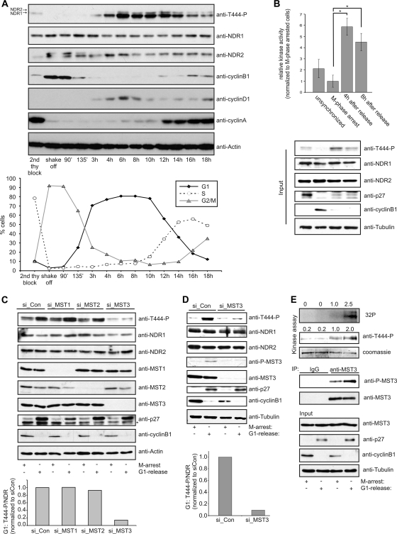
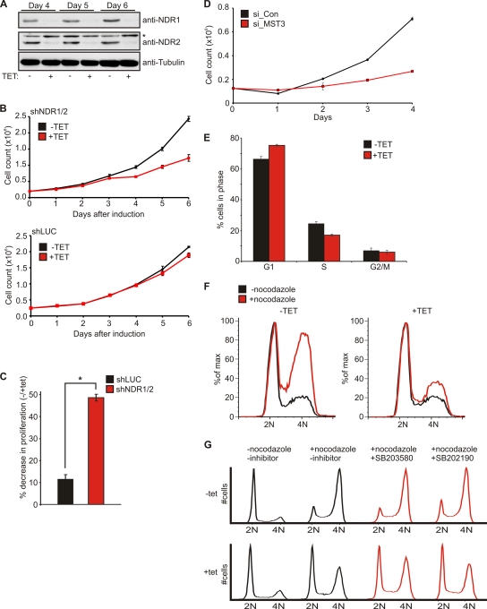
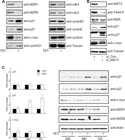
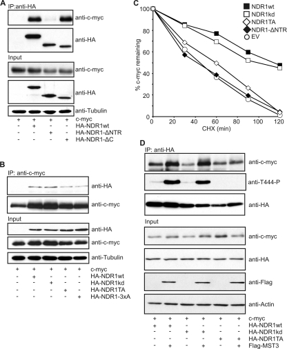
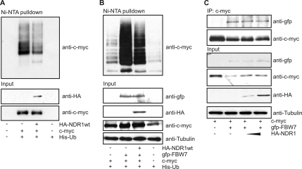
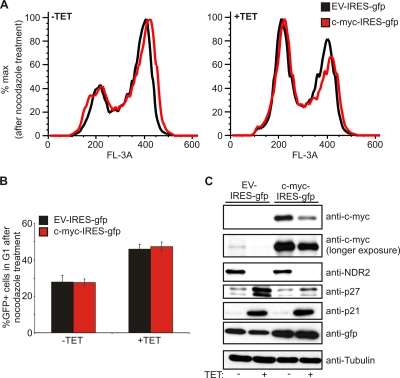
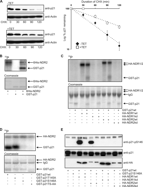
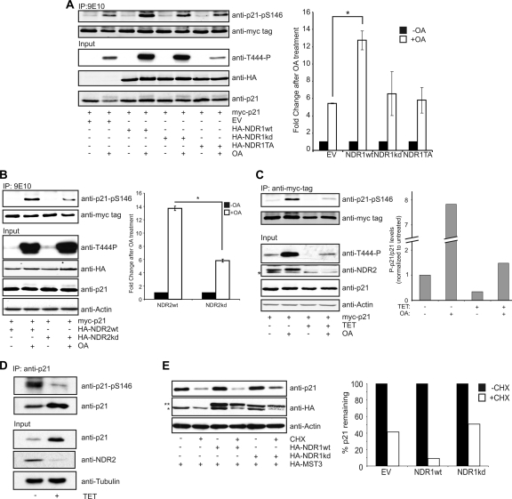
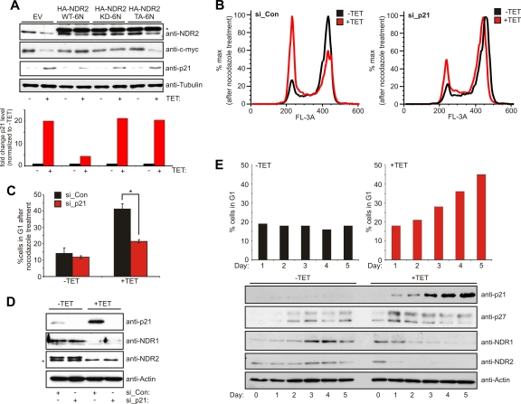
The G(1) phase of the cell cycle is an important integrator of internal and external cues, allowing a cell to decide whether to proliferate, differentiate, or die. Multiple protein kinases, among them the cyclin-dependent kinases (Cdks), control G(1)-phase progression and S-phase entry. With the regulation of apoptosis, centrosome duplication, and mitotic chromosome alignment downstream of the HIPPO pathway components MST1 and MST2, mammalian NDR kinases have been implicated to function in cell cycle-dependent processes. Although they are well characterized in terms of biochemical regulation and upstream signaling pathways, signaling mechanisms downstream of mammalian NDR kinases remain largely unknown. We identify here a role for human NDR in regulating the G(1)/S transition. In G(1) phase, NDR kinases are activated by a third MST kinase (MST3). Significantly, interfering with NDR and MST3 kinase expression results in G(1) arrest and subsequent proliferation defects. Furthermore, we describe the first downstream signaling mechanisms by which NDR kinases regulate cell cycle progression. Our findings suggest that NDR kinases control protein stability of the cyclin-Cdk inhibitor protein p21 by direct phosphorylation. These findings establish a novel MST3-NDR-p21 axis as an important regulator of G(1)/S progression of mammalian cells.


Figures

Fig. 1.
Fig. 1.
NDR kinases are activated by MST3 in the G1 phase of the cell cycle. (A) NDR kinases are activated in a cell cycle-dependent manner. Synchronized HeLa S3 cells were harvested after mitotic shake-off and replated in fresh medium for the indicated times. Activation of NDR1/2 was assessed using anti-T444-P, -NDR1, and -NDR2 antibodies. Cell cycle distribution was assessed using propidium iodide (PI) staining and FACS analysis. (B) Endogenous NDR kinase activity is increased in G1 phase. HeLa cells were arrested at the G2/M border using nocodazole treatment for 14 h and released for the indicated times before harvesting. Lysates were subjected to immunoblotting and immunoprecipitation of endogenous NDR species using a mixture of isoform-specific antibodies. NDR kinase activity was assessed using peptide kinase assays (n = 3; P < 0.002). (C) Depletion of MST1/2/3 kinases using isoform-specific siRNAs. HeLa cells were transfected with control siRNA (si_Con) or siRNAs targeting MST1/2/3 kinases (si_MST1, si_MST2, and si_MST3) and 48 h later were arrested with nocodazole for 14 h. Arrested cells were harvested or released into G1 for 8 h before harvesting. NDR activation was assessed using T444-P antibody. Cell cycle phases were confirmed by analyzing cyclin B1 and p27 expression (*, unspecific band). Phospho-T444 levels after G1 release were compared to those in control samples and analyzed using the Li-Cor Odyssey system. (D) Reduction of MST3 impairs G1 activation of NDR. HeLa cells were transfected with control siRNA (si_Con) or siRNA against MST3 (si_MST3) and treated and analyzed as described for panel C. MST3 activation was assessed using a P-MST4-T178/-MST3-T190/-STK25-T174-specific antibody (anti-P-MST3). Note that the P-MST3 signal disappears in the siMST3-treated samples. (E) Kinase activity of MST3 is increased in the G1 phase of the cell cycle. HeLa cells were arrested at the G2/M border using nocodazole treatment for 14 h and released for 8 h before harvesting. Lysates were subjected to immunoblotting and in parallel to immunoprecipitation (IP) of endogenous MST3 species using anti-MST3 or control antibodies. Endogenous MST3 kinase activity was assessed by an in vitro kinase assay using recombinant MBP-NDR1 K118R as a substrate. Reactions were analyzed by SDS-PAGE followed by immunoblotting against phosphorylation of Thr 444 in NDR1 and in parallel by exposure to a phosphorimager screen. NDR phosphorylation was quantified and normalized to the activity of MST3 in M phase.
Fig. 2.
Fig. 2.
shRNA-mediated knockdown of NDR1/2 results in cellular proliferation defects due to a G1 block. (A) Characterization of T-Rex-HeLa cells stably expressing shRNA against NDR1 and NDR2. Cells were seeded in 10-cm dishes, and shRNA expression was induced by the addition of tetracycline (TET) for the indicated times. Lysates from harvested cells were analyzed for NDR1 and NDR2 expression using isoform-specific antibodies (*, unspecific band). (B) NDR1/2 depletion results in proliferation defects. HeLa cells expressing shRNA against NDR1/2 (shNDR1/2) or firefly luciferase (shLUC) as a control were seeded in triplicates, and tetracycline was added to induce shRNA expression. After the indicated times, cells were harvested by trypsinization and counted using a ViCell automated cell counter. (C) Validation of proliferation defects in different clones stably expressing shNDR1/2 or shLUC (n = 3; P < 0.001). Experiments were performed as for panel B, and differences in proliferation were calculated as the percentage of cells without tetracycline to cells with tetracycline counted on day 6 after induction of shRNA expression. (D) Depletion of MST3 results in proliferation defects similar to those observed in NDR-depleted cells. HeLa cells were transfected with control siRNA (si_Con) or siRNA against MST3 (si_MST3); 24 h later, cells were seeded at defined densities in triplicates and cell counts were analyzed as for panel B. (E) Depletion of NDR1 and NDR2 results in an increase in G1 phase cells accompanied by a decrease in S phase cells. HeLa cells expressing shRNA against NDR1/2 were induced for 4 days with tetracycline. BrdU was added directly to the cell medium and left for 30 min before harvesting and processing for FACS analysis (n = 3). (F) Depletion of NDR1/2 results in G1 arrest. Knockdown of NDR1/2 was induced for 4 days using tetracycline. At 14 h before harvesting and processing for FACS analysis, cells were treated with 2.5 μg/ml nocodazole to induce G2/M accumulation. Fixed cells were stained with PI and analyzed by FACS. Histograms were overlaid to allow better comparison of cells in a given cell cycle phase. (G) Treatment of NDR1/2-depleted cells with inhibitors against p38 does not suppress G1 arrest. HeLa cells expressing shRNA against NDR1/2 were induced for 4 days with tetracycline. At 24 h before analysis, SB203580 or SB202190 (10 μM final concentration) was added to the cells. G1 arrest analysis was performed as for panel F.
Fig. 3.
Fig. 3.
Depletion of NDR1/2 kinases results in an increase in p21 and p27 protein levels and a decrease in c-myc protein level. (A) Reduced NDR kinase levels increase p21 and p27 and decrease c-myc. Knockdown of NDR1/2 was induced for 4 days, and cell lysates were analyzed for the expression of the indicated cell cycle regulators using Western blotting. (B) Depletion of MST3 increases p21 and p27 and decreases c-myc similarly to knockdown of NDR kinases. HeLa cells were transfected with control siRNA (si_Con) or siRNA against MST3 (si_MST3). After 3 days, cell lysates were analyzed by Western blotting (*, p27). (C) NDR1/2 depletion results in increased p27 mRNA levels, whereas changes in c-myc and p21 are observed only at the protein level. Knockdown of NDR1/2 was induced for the indicated times, and RNA extracts were prepared to analyze p21, p27, and c-myc mRNAs by quantitative RT-PCR. Values for p21, p27, and c-myc mRNAs are given as fold change relative to untreated samples (n = 3). In parallel, the samples were analyzed for the levels of the indicated proteins by Western blotting.
Fig. 4.
Fig. 4.
NDR1/2 in an active conformation stabilizes c-myc. (A) c-myc binds to the N-terminal region of NDR1. HEK293 cells were transfected with c-myc together with the indicated HA-tagged NDR1 constructs. NDR1 species were immunoprecipitated, and c-myc binding was analyzed by SDS-PAGE. (B) Binding of NDR1 to c-myc is modulated by hydrophobic motif phosphorylation (T444). HEK293 cells were transfected with the indicated NDR1 constructs (NDR1TA, T444A; NDR1-3xA, T74, S281, and T444A). c-myc was immunoprecipitated, and bound NDR1 species were analyzed by immunoblotting. (C) Overexpression of NDR1wt and NDR1kd stabilizes c-myc. HEK293 cells were transfected with c-myc and the indicated NDR1 cDNA; 24 h later, cells were treated with cycloheximide (CHX) for the indicated times and c-myc levels were analyzed using the Li-Cor Odyssey system. (D) Hydrophobic motif phosphorylation of NDR increases binding of NDR to c-myc. HEK293 cells were transfected with c-myc and the indicated NDR1 cDNAs together with a vector encoding FLAG-MST3, and complex formation was analyzed.
Fig. 5.
Fig. 5.
NDR overexpression impairs FBW7-mediated c-myc ubiquitination. (A) HEK293 cells were transfected with c-myc and His-tagged ubiquitin (His-Ub) together with HA-NDR1wt where indicated. Ubiquitinated proteins were pulled down from cell lysates using Ni-nitrilotriacetic acid (NTA)–Sepharose and analyzed by SDS-PAGE. (B) NDR decreases FBW7-mediated ubiquitination of c-myc. The experiment was performed as for panel A, but where indicated, GFP-FBW7 was coexpressed. (C) NDR1 does not compete with FBW7 for c-myc binding. HEK 293 cells were transfected with c-myc, FBW7, and increasing amounts of NDR1wt. c-myc was immunoprecipitated and analyzed for bound FBW7.
Fig. 6.
Fig. 6.
G1 arrest in NDR-depleted cells is not dependent on c-myc. (A) HeLa cells expressing shRNA against NDR1/2 were induced for 72 h with tetracycline and transfected with vectors expressing c-myc together with an IRES-GFP as a transfection marker. G1 arrest analysis was performed as described in the legend to Fig. 2F. Cell cycle profiles of GFP-positive cells were overlaid to allow for better comparison. (B) Analysis of GFP-positive cells in G1 after nocodazole arrest upon depletion of NDR1/2 and overexpression of c-myc (n = 3). (C) Analysis of c-myc, p21, and p27 in NDR1/2-depleted cells upon overexpression of c-myc. Cells were treated as described for panel A, but lysates were prepared before G1 arrest analysis.
Fig. 7.
Fig. 7.
NDR kinases phosphorylate p21 on S146 in vitro. (A) Depletion of NDR1/2 increases p21 stability. HeLa-shNDR1/2 cells were treated with tetracycline for 72 h. Prior to harvesting, cells were treated for the indicated times with CHX. p21 levels were analyzed using the Li-Cor Odyssey system (n = 3). p21 levels are depicted as the percentage remaining relative to the level at time zero, and trend lines were added to the data. r2 values for the trend lines are 0.98 (−TET) and 0.92 (+TET). Equation for the trend lines are y = 100e−0.0166x (−TET) and y = 100e−0.0083x (+TET). (B) Recombinant NDR2 phosphorylates recombinant p21. GST-p21 was used in an in vitro kinase assay with polyhistidine-tagged NDR2 from Sf9 cells in the presence of [γ-32P]ATP. After 30 min, reactions were analyzed by SDS-PAGE and exposure to a phosphorimager. (C) NDR1 and 2 phosphorylate p21 in vitro. HA-tagged NDR1/2wt or HA-NDR1/2kd was immunoprecipitated from okadaic acid-stimulated HEK293 cells and used for in vitro kinase assays with GST-p21 as a substrate. Reactions were analyzed as for panel B. (D) Ser 146 is the major site of p21 phosphorylated by NDR kinases. GST-p21 with mutated T145, S146, or T145/S146 phospho-acceptor sites was used as a substrate for in vitro kinase assays as described for panel B. (E) NDR1 and -2 phosphorylate p21 on Ser 146. GST-p21wt or GST-p21 S146A was used as a substrate in in vitro kinase assays using HA-tagged NDR1/2wt or HA-NDR1/2kd from okadaic acid-stimulated HEK 293 cells. Assays were analyzed by Western blotting using a phospho-specific antibody of p21 recognizing only if Ser 146 of p21 is phosphorylated.
Fig. 8.
Fig. 8.
NDR phosphorylates p21 in vivo and regulates p21 stability. (A) Overexpression of NDR1wt increases okadaic acid (OA)-induced phosphorylation of p21 on S146. HeLa cells were transfected with the indicated HA-tagged NDR1 species together with myc-tagged p21. Samples were stimulated with OA for 1 h before lysis where indicated. myc-tagged p21 was immunoprecipitated from lysates, and p21-pS146 levels were analyzed and quantified using the Li-Cor Odyssey system (n = 3; P < 0.002). p21-pS146 levels were normalized to controls without OA. (B) NDR2 phosphorylates p21 on S146 in vivo. HeLa cells were transfected with the HA-tagged NDR2wt or NDR2kd together with myc-tagged p21. Samples were stimulated with OA for 1 h before lysis where indicated. myc-tagged p21 was immunoprecipitated from lysates, and p21-pS146 levels were analyzed and quantified using the Li-Cor Odyssey system (n = 3; P < 0.0001). p21-pS146 levels were normalized to controls without OA. (C) Depletion of NDR1/2 decreases phosphorylation of p21 on S146. HeLa-shNDR1/2 cells were induced for 72 h with tetracycline and transfected with cDNA encoding myc-tagged p21. At 24 h after transfection, cells were stimulated with OA for 1 h and lysed. p21-pS146 levels were analyzed as for panel B (*, unspecific band). p21-pS146 levels were normalized to the untreated control sample. (D) Depletion of NDR results in a reduction of phosphorylated p21. HeLa-shNDR1/2 cells were induced for 72 h with tetracycline before lysis and immunoprecipitation of endogenous p21 were performed. p21-pS146 levels were analyzed by Western blotting. (E) Overexpression of NDR decreases p21 stability. HeLa cells were transfected with empty vector (EV) or with cDNAs encoding HA-NDR1wt or HA-NDR1kd in the presence of HA-MST3. Prior to lysis, cells were treated with CHX for 60 min where indicated (*, HA-MST3; **, HA-NDR).
Fig. 9.
Fig. 9.
G1 arrest after NDR kinase knockdown is rescued by reducing p21 levels. (A) The effects of NDR kinase depletion can be rescued depending on the NDR mutant. HeLa cells expressing shRNA against NDR2 were treated for 48 h with tetracycline and subsequently transfected with the indicated NDR2 mutants refractory to shNDR2 (*, HA-tagged NDR2). p21 levels were quantified using the Li-Cor Odyssey system. (B) HeLa cells expressing shRNA against NDR1/2 were treated with tetracycline for 48 h before being transfected with siRNA against p21 on two consecutive days. The cells were treated for G1 arrest analysis as described in the legend for Fig. 2F at 24 h after the second transfection. (C) Quantification of cells in G1 after nocodazole treatment (n = 3; P < 0.002). (D) Cells obtained from the experiment for panel B were analyzed for the expression of p21 and NDR1/2 (*, unspecific band). (E) p21 levels correlate with G1 arrest in NDR1/2-depleted cells. HeLa cells expressing shRNA against NDR1/2 were induced with tetracycline for the indicated times before harvest and lysis. Cells used for cell cycle analysis were treated with 2.5 μg/ml nocodazole for 14 h before harvest.

References

    1. Abbas T., Dutta A. 2009. p21 in cancer: intricate networks and multiple activities. Nat. Rev. Cancer 9:400–414 - PMC - PubMed
    1. Bichsel S. J., Tamaskovic R., Stegert M. R., Hemmings B. A. 2004. Mechanism of activation of NDR (nuclear Dbf2-related) protein kinase by the hMOB1 protein. J. Biol. Chem. 279:35228–35235 - PubMed
    1. Bidlingmaier S., Weiss E. L., Seidel C., Drubin D. G., Snyder M. 2001. The Cbk1p pathway is important for polarized cell growth and cell separation in Saccharomyces cerevisiae. Mol. Cell. Biol. 21:2449–2462 - PMC - PubMed
    1. Bothos J., Tuttle R. L., Ottey M., Luca F. C., Halazonetis T. D. 2005. Human LATS1 is a mitotic exit network kinase. Cancer Res. 65:6568–6575 - PubMed
    1. Chen C. B., Ng J. K., Choo P. H., Wu W., Porter A. G. 2009. Mammalian sterile 20-like kinase 3 (MST3) mediates oxidative-stress-induced cell death by modulating JNK activation. Biosci. Rep. 29:405–415 - PubMed

Publication types

MeSH terms