. 2011 Feb 8;108(6):2605-10.
doi: 10.1073/pnas.1015788108. Epub 2011 Jan 24.

Optical probing of a dynamic membrane interaction that regulates the TREK1 channel

Affiliations

Optical probing of a dynamic membrane interaction that regulates the TREK1 channel

Guillaume Sandoz et al. Proc Natl Acad Sci U S A. .

Abstract

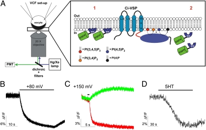
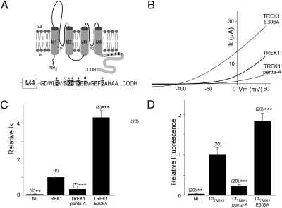
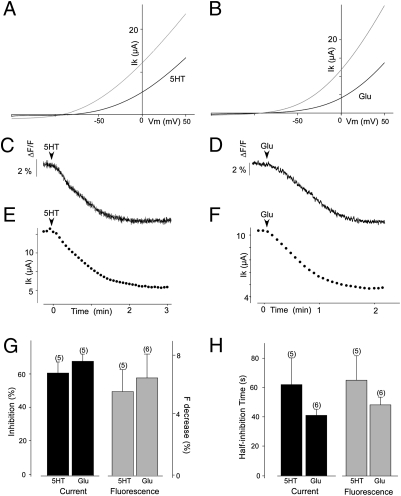
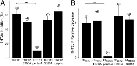
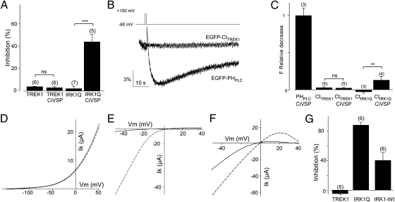
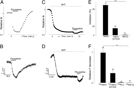
TREK channels produce background currents that regulate cell excitability. These channels are sensitive to a wide variety of stimuli including polyunsaturated fatty acids (PUFAs), phospholipids, mechanical stretch, and intracellular acidification. They are inhibited by neurotransmitters, hormones, and pharmacological agents such as the antidepressant fluoxetine. TREK1 knockout mice have impaired PUFA-mediated neuroprotection to ischemia, reduced sensitivity to volatile anesthetics, altered perception of pain, and a depression-resistant phenotype. Here, we investigate TREK1 regulation by Gq-coupled receptors (GqPCR) and phospholipids. Several reports indicate that the C-terminal domain of TREK1 is a key regulatory domain. We developed a fluorescent-based technique that monitors the plasma membrane association of the C terminus of TREK1 in real time. Our fluorescence and functional experiments link the modulation of TREK1 channel function by internal pH, phospholipid, and GqPCRs to TREK1-C-terminal domain association to the plasma membrane, where increased association results in greater activity. In keeping with this relation, inhibition of TREK1 current by fluoxetine is found to be accompanied by dissociation of the C-terminal domain from the membrane.


Conflict of interest statement

The authors declare no conflict of interest.

Figures

Fig. 1.
Fig. 1.
Development of cell-based assay to study cell membrane binding of GFP-fusion proteins. (A) Measuring membrane binding of GFP protein. Xenopus oocytes expressing the protein of interest are voltage clamped. The fluorescence of the EGFP-fusion protein is recorded by epifluorescence (1). The cortical layer of pigment granules in the animal hemisphere and the opaque cytoplasmic compartment provide a mask for cytoplasmic fluorescence (2). When the fusion proteins lose their interaction with the plasma membrane, the fusion proteins diffuse into the cell, inducing a decrease of fluorescence. (BD) PH-PLC domain assay. Representative example of EFGP-PHPLC fluorescence decrease induced by Ci-VSP activation (30-s pulse from −80 to +80 mV) (D) and 5HT2cR activation (C) in Xenopus oocyte. (C) Representative example of tagRFP-PHPLC (red) fluorescence decrease and EFGP-PHOSH1 fluorescence increase (green) induced by Ci-VSP activation (200-ms pulse from −80 to +150 mV).
Fig. 2.
Fig. 2.
The current amplitude of TREK1 mutants are correlated with the membrane fluorescence of the corresponding EGFP-CtTREK1 mutants. (A) Membrane topology of TREK1. The sequence of the cytoplasmic C terminus of TREK1 starting at Gly293 is indicated. +, basic residues (R297, K301, K302, K304, and R311); open circles, PKC (S300) phosphorylation site; and dark circle, pH sensor site (E306). (B) Currents were elicited by voltage ramps (from −150 to +50 mV, 1 s in duration). (C) Normalized amplitude of noninjected oocytes (NI), TREK1-WT, TREK1-Penta-A, and TREK1-E306. (D) Normalized membrane fluorescence level of oocytes expressing EGFP-CtTREK1, EGFP-CtTREK1-penta-A, and EGFP-CtTREK1-E306A. Student's t test (**P < 0.01, ***P < 0.001) show the difference between TREK1 and TREK1 mutants (or EGFP-CtTREK1 and EGFP-CtTREK1 mutants). The numbers of cells tested are indicated in parentheses.
Fig. 3.
Fig. 3.
Gq-coupled receptor activation regulates TREK1 currents and membrane association of EGFP-CtTREK1 constructs. (A and B) Effect of activation of 5HTR2bR (A) or mGluRI on TREK1 expressed in Xenopus oocytes. Currents were elicited by voltage ramp (from −150 to +50 mV, 1 s in duration). (C and D) Representative example of EGFP-CtTREK1 fluorescence decrease induced by 5HT2cR (C) and mGluRI (D) in oocytes. (E and F) Representative example of TREK1 current inhibition induced by 5HT2cR (E) and mGluRI (F) in oocytes. Effect of 5HT2cR and mGluRI activation on TREK1 current and on membrane fluorescence of EGFP-CtTREK1. (G) Percentage of inhibition (black) or of fluorescence decrease (gray) induced by 5HT2cR and mGluRI activation. Inhibitions and fluorescence decreases are statistically significant (P < 0.001) for both 5HT2cR and mGluRI. (H) Half current inhibition time (black) or the half decrease time of fluorescence induced by 5HT2cR and mGluRI activation (gray). The numbers of cells tested are indicated in parentheses.
Fig. 4.
Fig. 4.
Point mutations in C-terminal domain regulate TREK1 response to GPCR activation. (A) Percentage of current inhibition by 5HT2cR activation for TREK1-WT, TREK1-Penta-A, TREK1-E306, TREK1-S300A, and TREK1-WT preincubated with calphostatin C. (B) Relative fluorescence decrease induced by 5HT2cR activation for EGFP-CtTREK1 WT, EGFP-CtTREK1-Penta-A, EGFP-CtTREK1-E306, EGFP-CtTREK1-S300A, and TREK1-WT preincubated with calphostatin C (overnight 5 μM). The asterisks indicate the significance (P < 0.05) of the difference between TREK1 mutants and TREK1-WT (or EGFP-CtTREK1 mutants and EGFP-CtTREK1). Student's t test: ***P < 0.001. The numbers of cells tested are indicated in parentheses.
Fig. 5.
Fig. 5.
TREK1 is not directly regulated by PI(4,5)P2. (A) TREK1 current inhibition induced by the activation of Ci-VSP by a prepulse to +150 mV (200 ms). Currents were measured 15 s after the prepulse. (B) Representative examples of EGFP-CtTREK1 and EFGP-PHPLC fluorescence decrease induced by Ci-VSP activation (pulse at +150 mV from −80 mV, 200-ms duration). (C) Relative fluorescence decrease of EFGP-PHPLC and EGFP-CtTREK1 and EGFP-CtIRK-R228Q induced by Ci-VSP. (DG) PI(4,5)P2 depletion by rapamycin-inducible 5-phosphatase [PI(4,5)P2→ PI(4)P] failed to regulate TREK current. (DF) Effect of activation of 5-phosphatase by rapamycin (5 μM) on TREK1 (D), IRK1-R228Q (E), and IRK1-WT (F). Currents before (dashed lines) and after rapamycin (solid lines) were elicited by voltage ramps (from −50 to +50 mV, 1 s in duration). (G) Inhibition induced by PI(4,5)P2 depletion by the rapamycin system on TREK1, IRK1-R228Q, and IRK1-WT. Student's t test: **P < 0.01, ***P < 0.001. The numbers of cells tested are indicated in parentheses.
Fig. 6.
Fig. 6.
Fluoxetine reduces TREK1 current and induces EGFP-CtTREK1 dissociation from the membrane. (A and C) Effect of fluoxetine application (100 μM) on TREK1 current and on the membrane fluorescence of EGFP-CtTREK1, respectively. (B and D) Effect of fluoxetine application (100 μM) on TREK1 current and on the membrane fluorescence of EGFP-CtTREK1 after 5HT2cR activation. (E) The histograms represent current inhibition induced by fluoxetine for TREK1 before and after 5HT2cR activation and for IRK1. (F) Relative fluorescence decrease induced by fluoxetine for TREK1 before and after 5HT2cR activation and for EFGP-PHPLC and and EGFP-CtIRK1Q. Student's t test: ***P < 0.001. The numbers of cells tested are indicated in parentheses.

References

    1. Fink M, et al. Cloning, functional expression and brain localization of a novel unconventional outward rectifier K+ channel. EMBO J. 1996;15:6854–6862. - PMC - PubMed
    1. Maingret F, et al. TREK-1 is a heat-activated background K(+) channel. EMBO J. 2000;19:2483–2491. - PMC - PubMed
    1. Patel AJ, et al. A mammalian two pore domain mechano-gated S-like K+ channel. EMBO J. 1998;17:4283–4290. - PMC - PubMed
    1. Sandoz G, Douguet D, Chatelain F, Lazdunski M, Lesage F. Extracellular acidification exerts opposite actions on TREK1 and TREK2 potassium channels via a single conserved histidine residue. Proc Natl Acad Sci USA. 2009;106:14628–14633. - PMC - PubMed
    1. Maingret F, Patel AJ, Lesage F, Lazdunski M, Honoré E. Mechano- or acid stimulation, two interactive modes of activation of the TREK-1 potassium channel. J Biol Chem. 1999;274:26691–26696. - PubMed

Publication types

MeSH terms

Substances

LinkOut - more resources