. 2011 Feb 8;108(6):2534-9.
doi: 10.1073/pnas.1019577108. Epub 2011 Jan 24.

The cell-adhesion G protein-coupled receptor BAI3 is a high-affinity receptor for C1q-like proteins

Affiliations

The cell-adhesion G protein-coupled receptor BAI3 is a high-affinity receptor for C1q-like proteins

Marc F Bolliger et al. Proc Natl Acad Sci U S A. .

Abstract

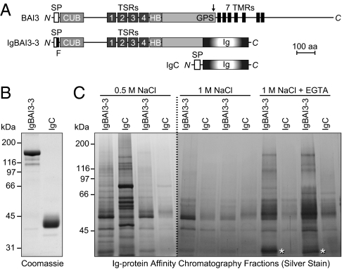
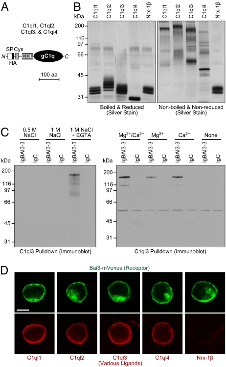
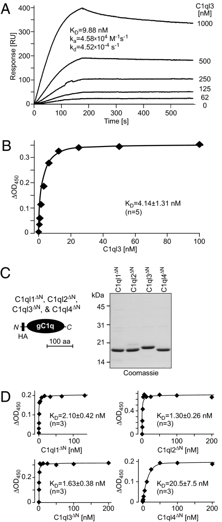
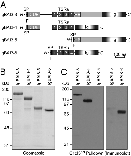
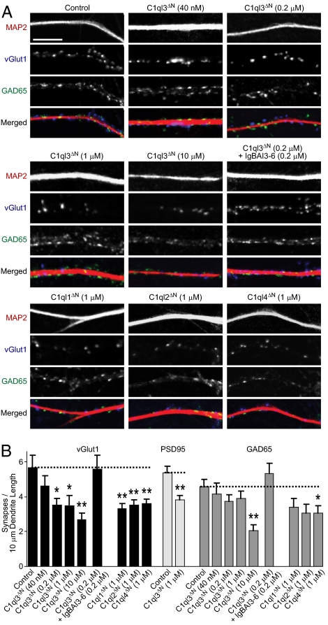
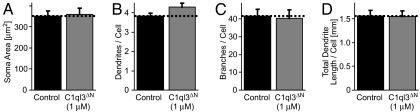
C1q-like genes (C1ql1-C1ql4) encode small, secreted proteins that are expressed in differential patterns in the brain but whose receptors and functions remain unknown. BAI3 protein, in contrast, is a member of the cell-adhesion class of G protein-coupled receptors that are expressed at high levels in the brain but whose ligands have thus far escaped identification. Using a biochemical approach, we show that all four C1ql proteins bind to the extracellular thrombospondin-repeat domain of BAI3 with high affinity, and that this binding is mediated by the globular C1q domains of the C1ql proteins. Moreover, we demonstrate that addition of submicromolar concentrations of C1ql proteins to cultured neurons causes a significant decrease in synapse density, and that this decrease was prevented by simultaneous addition of the thrombospondin-repeat fragment of BAI3, which binds to C1ql proteins. Our data suggest that C1ql proteins are secreted signaling molecules that bind to BAI3 and act, at least in part, to regulate synapse formation and/or maintenance.


Conflict of interest statement

The authors declare no conflict of interest.

Figures

Fig. 1.
Fig. 1.
Identification of C1ql proteins as BAI3 ligands. (A) Schematic depiction of the domain organization of BAI3 (1,522 amino acids), of the BAI3 Ig-fusion protein IgBAI3-3, and of the control Ig-fusion protein IgC. SP, signal peptide; F, FLAG tag; CUB, complement C1r/C1s-Uegf-Bmp1 domain; TSRs, thrombospondin repeats; HB, hormone-binding domain; GPS, GPCR proteolysis sequence (proteolytic site denoted by an arrow); 7 TMR, seven-transmembrane region; Ig, Fc domain of human IgG. (B) Coomassie blue–stained SDS gel of Ig-fusion proteins produced in HEK293 cells and purified by protein A–Sepharose affinity chromatography. (C) Silver-stained SDS gel of a representative pull-down experiment. Mouse brain homogenates were incubated with protein A–Sepharose beads containing either IgBAI3-3 or IgC. After the beads were washed, bound proteins were sequentially eluted with buffers containing 0.5 M NaCl, 1 M NaCl, and 1 M NaCl with 5 mM EGTA. Two fractions per buffer were collected and analyzed. Mass spectrometry analysis identified C1ql2 and C1ql3 in the band marked by an asterisk.
Fig. 2.
Fig. 2.
Characterization of BAI3 binding by C1ql proteins. (A) Domain structure of C1ql proteins C1ql1–C1ql4 (238–287 amino acids). SP, signal peptide; Cys, cysteine loop domain; COL, collagen-like domain; gC1q, globular C1q domain. An HA epitope was inserted immediately following the signal peptide. (B) Silver-stained SDS gel of purified HA-tagged full-length C1ql proteins and of a recombinant protein containing the extracellular neurexin-1β sequences loaded onto SDS gels with (Left) or without (Right) reducing agents and boiling. Proteins were expressed in HEK293 cells and purified from the medium by anti-HA agarose. (C) Analyses of C1ql3-mediated pull-downs of recombinant IgBAI3-3 protein, using IgC as a negative control. HA-tagged C1ql3 immobilized on an anti-HA column was incubated with IgBAI3-3 or IgC, and bound proteins were analyzed by immunoblot analysis for human Ig. (Left) Eluates obtained after binding in Ca2+/Mg2+-containing buffer and elution with the indicated solutions. (Right) Proteins bound to immobilized C1ql3 in buffers containing the indicated divalent cations. (D) Cell-surface labeling assay demonstrating that all four C1ql isoforms bind to BAI3. HEK293 cells transfected with a vector encoding mVenus-tagged BAI3 (green) were incubated with purified HA-tagged C1ql1–C1ql4 and neurexin-1β (extracellular sequences). Bound proteins were visualized by immunofluorescence labeling (red). (Scale bar: 10 μm.)
Fig. 3.
Fig. 3.
Nanomolar affinity of C1ql1, C1ql2, C1ql3, and C1ql4 for BAI3. (A) Surface plasmon resonance measurements. Full-length C1ql3 was injected at the indicated concentrations onto chips containing immobilized IgBAI3-3 or IgC for 180 s, followed by injection of buffer alone. Deduced binding constants are listed in the graph. RU, resonance units. (B) Cell-surface binding measurements with full-length C1ql3 as a ligand. HEK293 cells were transfected with a BAI3 cDNA or mock-transfected, incubated with purified HA-tagged full-length C1ql3 at the indicated concentrations, and fixed. Bound protein was detected with a peroxidase-conjugated antibody against the HA epitope. The signal shown was subtracted for background observed with mock-transfected cells. Data are from a representative experiment independently repeated five times, with Scatchard fits resulting in the indicated binding constant (mean ± SEM). (C) Schematic diagram (Left) and Coomassie blue–stained SDS gel (Right) of recombinant gC1q domains of all four C1ql proteins. gC1q domains were expressed with an N-terminal HA tag as GST-fusion proteins in Escherichia coli, and GST was removed by thrombin cleavage after purification with glutathione-Sepharose. gC1q domains are referred to as C1ql1ΔN–C1ql4ΔN because the N-terminal cysteine sequence and collagen domain were deleted from the C1ql proteins to generate these recombinant domains. (D) Binding curves generated as described in B, but with HA-tagged N-terminally truncated C1ql molecules consisting only of the HA-tagged gC1q domains of the respective proteins. Data shown are from a representative experiment independently repeated three times, with Scatchard fits resulting in the indicated binding constants (mean ± SEM).
Fig. 4.
Fig. 4.
BAI3 fragment containing thrombospondin repeats binds to the gC1q domain of C1ql3. (A) Schematic diagram of BAI3 deletion constructs. Abbreviations are as in Fig. 1. (B) Coomassie blue–stained SDS gels of BAI3 Ig-fusion proteins expressed in HEK293 cells and purified from the medium by anti-FLAG agarose. (C) Pull-down experiments with the HA-tagged gC1q domain of C1ql3 that was immobilized on an HA-antibody column and used as an affinity matrix for the indicated purified BAI3 Ig-fusion proteins. Bound proteins were detected by immunoblot analysis for the FLAG epitopes. Data show representative experiments independently repeated three times.
Fig. 5.
Fig. 5.
Incubation of primary hippocampal neurons with recombinant gC1q domains from C1ql1–C1ql4 decreases synapse density. (A) Representative images of dendrites from cultured hippocampal neurons that were incubated with recombinant HA-tagged gC1q domains of the indicated C1ql proteins at DIV10 and analyzed at DIV14 by immunofluorescence staining with antibodies to MAP2 (to label dendrites) and vGlut1 or GAD65 as markers for excitatory and inhibitory synapses, respectively. (Scale bar: 10 μm.) (B) Summary graphs of excitatory and inhibitory synapse densities measured by staining with antibodies to vGlut1 and PSD95 (for excitatory synapses) or to GAD65 (for inhibitory synapses). Synapse densities were determined in neurons treated as described in A, with the incubation conditions indicated below each bar. Graphs show mean ± SEM of three independent experiments (two-tailed Student's t test; *P < 0.05; **P < 0.01).
Fig. 6.
Fig. 6.
Treatment of cultured hippocampal neurons with the C1ql3 gC1q domain does not alter overall neuron size, dendrite length, or dendritic branching. Hippocampal neurons were treated as described in Fig. 5 with recombinant C1ql3 gC1q domain (1 μM). Graphs show the mean size of the neuronal soma (A), number of dendritic processes per soma (B), number of dendritic branches per cell (C), and total length of dendrites per cell (D). Data are mean ± SEM of three independent experiments.

References

    1. Yona S, Lin HH, Siu WO, Gordon S, Stacey M. Adhesion-GPCRs: Emerging roles for novel receptors. Trends Biochem Sci. 2008;33:491–500. - PubMed
    1. Lagerström MC, Schiöth HB. Structural diversity of G protein-coupled receptors and significance for drug discovery. Nat Rev Drug Discov. 2008;7:339–357. - PubMed
    1. Bjarnadóttir TK, et al. The human and mouse repertoire of the adhesion family of G protein-coupled receptors. Genomics. 2004;84:23–33. - PubMed
    1. Krasnoperov V, et al. Post-translational proteolytic processing of the calcium-independent receptor of alpha-latrotoxin (CIRL), a natural chimera of the cell adhesion protein and the G protein-coupled receptor: Role of the G protein-coupled receptor proteolysis site (GPS) motif. J Biol Chem. 2002;277:46518–46526. - PubMed
    1. Lin HH, et al. Autocatalytic cleavage of the EMR2 receptor occurs at a conserved G protein-coupled receptor proteolytic site motif. J Biol Chem. 2004;279:31823–31832. - PubMed

Publication types