. 2011 Feb 8;108(6):2205-9.
doi: 10.1073/pnas.1019381108. Epub 2011 Jan 24.

Casein kinase-2 mediates cell survival through phosphorylation and degradation of inositol hexakisphosphate kinase-2

Affiliations

Casein kinase-2 mediates cell survival through phosphorylation and degradation of inositol hexakisphosphate kinase-2

Anutosh Chakraborty et al. Proc Natl Acad Sci U S A. .

Abstract

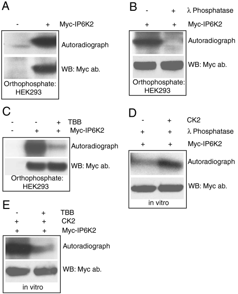
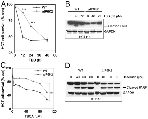
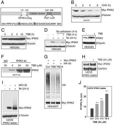
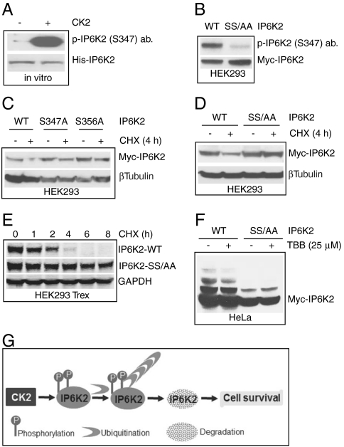
The inositol pyrophosphate, diphosphoinositol pentakisphosphate, regulates p53 and protein kinase Akt signaling, and its aberrant increase in cells has been implicated in apoptosis and insulin resistance. Inositol hexakisphosphate kinase-2 (IP6K2), one of the major inositol pyrophosphate synthesizing enzymes, mediates p53-linked apoptotic cell death. Casein kinase-2 (CK2) promotes cell survival and is upregulated in tumors. We show that CK2 mediated cell survival involves IP6K2 destabilization. CK2 physiologically phosphorylates IP6K2 at amino acid residues S347 and S356 contained within a PEST sequence, a consensus site for ubiquitination. HCT116 cells depleted of IP6K2 are resistant to cell death elicited by CK2 inhibitors. CK2 phosphorylation at the degradation motif of IP6K2 enhances its ubiquitination and subsequent degradation. IP6K2 mutants at the CK2 sites that are resistant to CK2 phosphorylation are metabolically stable.


Conflict of interest statement

The authors declare no conflict of interest.

Figures

Fig. 1.
Fig. 1.
CK2 phosphorylates IP6K2 A. Myc-IP6K2 phosphorylation in HEK293 cells detected by orthophosphate labeling. B. Specificity of IP6K2 phosphorylation is confirmed by λ phosphatase treatment of the radiolabeled IP6K2 protein after orthophosphate labeling. C. TBB treatment (50 μM, 3 h) abolishes Myc-IP6K2 phosphorylation in HEK293 cells. D. CK2 phosphorylation of immunoprecipitated and λ phosphatase treated Myc-IP6K2 in vitro. E. TBB (5 μM) inhibits CK2 phosphorylation of Myc-IP6K2 in vitro.
Fig. 2.
Fig. 2.
IP6K2 mediates cell death associated with CK2 inhibition A. IP6K2Δ HCT116 cells display resistance to inhibitory effect of TBB on cell survival compared to WT HCT cells. (***p < 0.001). B. TBB mediated PARP cleavage is absent in IP6K2Δ HCT116 cells. C. TBCA’s inhibitory effect on cell survival is greatly reduced in IP6K2Δ HCT116 cells. (***p < 0.001). D. Resorufin induced apoptosis, as indicated by PARP cleavage, is reduced in IP6K2Δ HCT116 cells.
Fig. 3.
Fig. 3.
IP6K2 stability is enhanced by CK2 inhibition A. CK2 phosphorylation sites at the degradation specific PEST motif of IP6K2. B. Myc-IP6K2 protein displays a half-life of 2 h as measured by cycloheximide treatment of HEK293 cells. C. TBB (50 μM) stabilizes Myc-IP6K2 protein levels in a time-dependent fashion. D. TBB (50 μM) protects tetracycline removal induced IP6K2 protein degradation. E. TBB (50 μM, 6 h) stabilizes endogenous IP6K2 levels in HEK293 cells. F. Immunoprecipitation of endogenous IP6K2 from HCT116 cells reveals increased protein levels after TBB treatment. G. IP6K2 ubiquitination is significantly reduced after TBB (50 μM) or TBCA (10 μM) treatment, H. TBB stabilizes Myc-IP6K2 in U2OS cells. I. The MG132 treatment reveals multiple ubiquitinated Myc-IP6K2. J. TBB increases IP7 levels in U2OS cells stably expressing Myc-IP6K2. (***p < 0.001).
Fig. 4.
Fig. 4.
IP6K2 destabilization is associated with CK2 phosphorylation at serines 347 and 356 A. CK2 phosphorylation of bacterially purified His-tagged IP6K2 is detected by α-pIP6K2 (S347) antibody. Total protein was detected by coommassie blue staining. B. Myc-IP6K2 S347/356AA double mutant is not recognized by the α-pIP6K2 (S347) antibody. WT and mutant IP6K2 were overexpressed in HEK293 cells and were immunoprecipitated using α-Myc antibody. C. IP6K2 S347A and S356A single mutants display partial but not total stability after cycloheximide treatment. D. IP6K2 S347/356AA double mutant does not degrade after 4 h cycloheximide treatment. E. Substantial increase in half-life of IP6K2 S347/356AA double mutant. The half-life of the mutant is 8 h as compared to 2 h for WT IP6K2. F.TBB decreases ubiquitination of WT-IP6K2, whereas the mutant is resistant to ubiquitination and hence unaffected by TBB treatment. G. Model showing CK2 phosphorylation mediated ubiquitination and degradation of IP6K2.

References

    1. Berridge MJ, Lipp P, Bootman MD. The versatility and universality of calcium signalling. Nat Rev Mol Cell Biol. 2000;1:11–21. - PubMed
    1. Hand CE, Honek JF. Phosphate transfer from inositol pyrophosphates InsP5PP and InsP4(PP)2: a semi-empirical investigation. Bioorg Med Chem Lett. 2007;17:183–188. - PubMed
    1. Barker CJ, Illies C, Gaboardi GC, Berggren PO. Inositol pyrophosphates: structure, enzymology and function. Cell Mol Life Sci. 2009;66:3851–3871. - PMC - PubMed
    1. Shears SB. Diphosphoinositol polyphosphates: metabolic messengers? Mol Pharmacol. 2009;76:236–252. - PMC - PubMed
    1. Mulugu S, et al. A conserved family of enzymes that phosphorylate inositol hexakisphosphate. Science. 2007;316:106–109. - PubMed

Publication types

Substances