. 2011 Apr 1;286(13):11696-706.
doi: 10.1074/jbc.M110.206623. Epub 2011 Jan 24.

Peroxiredoxin 6 phosphorylation and subsequent phospholipase A2 activity are required for agonist-mediated activation of NADPH oxidase in mouse pulmonary microvascular endothelium and alveolar macrophages

Affiliations

Peroxiredoxin 6 phosphorylation and subsequent phospholipase A2 activity are required for agonist-mediated activation of NADPH oxidase in mouse pulmonary microvascular endothelium and alveolar macrophages

Shampa Chatterjee et al. J Biol Chem. .

Abstract

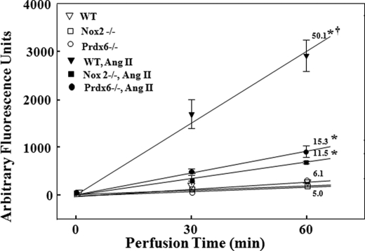
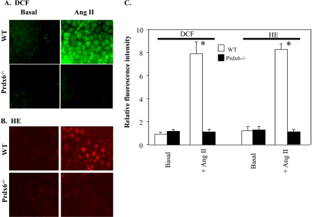
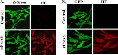
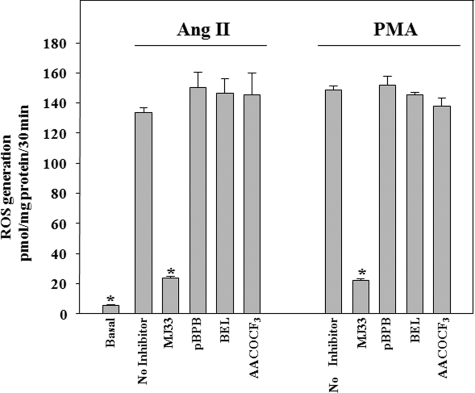
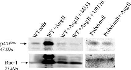
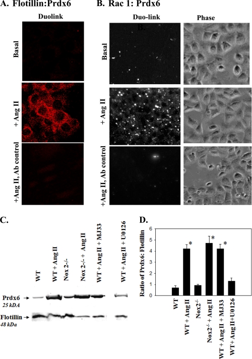
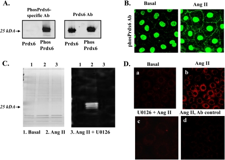
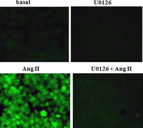
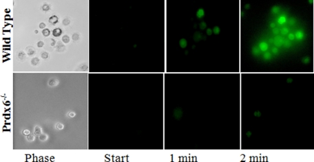
Peroxiredoxin 6 (Prdx6), a bifunctional enzyme with glutathione peroxidase and phospholipase A2 (PLA(2)) activities, participates in the activation of NADPH oxidase 2 (NOX2) in neutrophils, but the mechanism for this effect is not known. We now demonstrate that Prdx6 is required for agonist-induced NOX2 activation in pulmonary microvascular endothelial cells (PMVEC) and that the effect requires the PLA(2) activity of Prdx6. Generation of reactive oxygen species (ROS) in response to angiotensin II (Ang II) or phorbol 12-myristate 13-acetate was markedly reduced in perfused lungs and isolated PMVEC from Prdx6 null mice. Rac1 and p47(phox), cytosolic components of NOX2, translocated to the endothelial cell membrane after Ang II treatment in wild-type but not Prdx6 null PMVEC. MJ33, an inhibitor of Prdx6 PLA(2) activity, blocked agonist-induced PLA(2) activity and ROS generation in PMVEC by >80%, whereas inhibitors of other PLA(2)s were ineffective. Transfection of Prx6 null cells with wild-type and C47S mutant Prdx6, but not with mutants of the PLA(2) active site (S32A, H26A, and D140A), "rescued" Ang II-induced PLA(2) activity and ROS generation. Ang II treatment of wild-type cells resulted in phosphorylation of Prdx6 and its subsequent translocation from the cytosol to the cell membrane. Phosphorylation as well as PLA(2) activity and ROS generation were markedly reduced by the MAPK inhibitor, U0126. Thus, agonist-induced MAPK activation leads to Prdx6 phosphorylation and translocation to the cell membrane, where its PLA(2) activity facilitates assembly of the NOX2 complex and activation of the oxidase.


Figures

FIGURE 1.
FIGURE 1.
ROS production following Ang II stimulation is markedly decreased in isolated perfused mouse lungs from Prdx6 null mice. Lungs from WT, Prdx6 null, and NOX2 null were isolated and perfused in a recirculating system containing Amplex Red (25 μm) and HRP (25 μg/ml). ROS generation was measured in arbitrary fluorescence units by Amplex Red oxidation at 30 and 60 min of perfusion. Ang II when present was 50 μm. The numbers above or below the lines indicate the slope (change in arbitrary fluorescence units/min) calculated by least mean squares. *, p < 0.05 versus the corresponding control (no Ang II); †, p < 0.05 versus other Ang II-stimulated conditions. Error bars, S.E.
FIGURE 2.
FIGURE 2.
Prdx6 is required for ROS production by PMVEC in response to Ang II treatment. PMVEC derived from WT or Prdx6 null mice were monitored by fluorescence microscopy for ROS production with and without the addition of Ang II (10 μm) by dihydrochlorofluorescein oxidation to DCF (green) (A) or by HE oxidation (red) (B). C, to quantify fluorescence intensity, 8–10 PMVEC in each of five fields were randomly selected in the phase images, and then fluorescence of these cells in arbitrary units was quantified using Metamorph software. Results are expressed as mean ± S.E. (error bars) for n = 8. *, p < 0.05 versus basal (no Ang II).
FIGURE 3.
FIGURE 3.
Transfection of Prdx6 null mouse PMVEC with a vector expressing Prdx6 restores Ang II-induced ROS generation. PMVEC isolated from Prdx6 null mice were transfected with vector only or vector expressing either pIRES2-ZsGreen1 vector (Control) or mouse Prdx6:pIRES2-ZsGreen1 (mPrdx6) (A) or pGFP-C1 (Control) or rat Prdx6:GFP-C1 (rPrdx6) (B). Cells were evaluated by fluorescence microscopy. Green fluorescence (ZsGreen or GFP) indicates transfection. Transfected cells were treated with Ang II (10 μm) for 30 min and monitored for ROS generation (red) by oxidation of HE.
FIGURE 4.
FIGURE 4.
ROS generation with Ang II or PMA treatment is blocked by an inhibitor of Prdx6 PLA2 activity but not by other PLA2 inhibitors. PMVEC were pretreated with inhibitor for 10 min and then incubated with Ang II (10 μm) or PMA (10 nm). ROS production was measured during the incubation with agonist by the Amplex Red oxidation method. Inhibitors were MJ33 (1 mol % in liposomes), pBPB (20 μm), BEL (100 μm), and AACOCF3 (100 μm). Values are mean ± S.E. (error bars) for n = 3. *, p < 0.05 versus Ang II or PMA alone. Basal (no additions) was the same for both Ang II and PMA.
FIGURE 5.
FIGURE 5.
NOX2 cytosolic components (Rac1 and p47phox) translocate to the cell membrane upon Ang II treatment in wild-type but not Prdx6 null PMVEC. PMVEC from WT or Prdx6 null mice were treated for 30 min with 10 μm Ang II. Where indicated, wild-type cells also were pretreated with MJ33 (1 mol % in liposomes) or U0126 (10 μm) for 30 min before the addition of Ang II. Endothelial cell membranes were isolated by subcellular fractionation and immunoblotted using antibodies to Rac1 or p47phox. Rac1 appears as a doublet as described previously (41); the lower band was used for quantitation (Table 6). Results were obtained with separate gels for wild-type and Prdx6 null cells. Gels were cut and processed for two different proteins as described under “Experimental Procedures.”
FIGURE 6.
FIGURE 6.
Prdx6 associates with the plasma membrane upon Ang II treatment. Duolink procedure to evaluate co-localization of Prdx6 in PMVEC with flotillin (A), an integral plasma membrane protein, and Rac1 (B), a cytoplasmic protein that translocates to the plasma membrane upon stimulation. Fluorescence indicates proximity (<40 nm) of the two proteins; phase images in B show cell density. Upper panels, basal (no Ang II); middle and lower panels, Ang II (10 μm). Upper and middle panels were treated with anti-Prdx6 (polyclonal Ab) and either anti-flotillin or anti-Rac1 (mAb); lower panels are the Ab control (IgG from rabbit and mouse). Images were obtained similarly for A and B, but positive results are shown as red fluorescence (A) or converted to black and white (B). The phase image is shown in B because the cell borders are not apparent in the fluorescence image. C, immunoblots of the cell membrane fraction isolated by gradient centrifugation from mouse WT and NOX2 null PMVEC with and without Ang II (10 μm) treatment using anti-Prdx6 and anti-flotillin antibodies. MJ33 (1 mol % in liposomes) was used as an inhibitor of Prdx6 PLA2 activity, and U0126 (10 μm) was used as a MAPK inhibitor. The gap indicates where several non-relevant lanes were removed. The blot was cut horizontally for simultaneous analysis of both proteins as described under “Experimental Procedures.” D, densitometric quantitation (mean ± S.E. (error bars) for n = 3) of the bands on immunoblot in arbitrary units using ImageJ software; results are expressed as the ratio of Prdx6 to flotillin. *, p < 0.05 versus wild-type control (no Ang II).
FIGURE 7.
FIGURE 7.
Prdx6 phosphorylation upon Ang II treatment. A, immunoblot to show specificity of the polyclonal Ab to phosphorylated Prdx6. Recombinant mouse Prdx6 was phosphorylated in vitro by incubation with ERK2 MAPK. The Ab to the phosphorylated peptide reacts with phosphorylated Prdx6 but does not recognize non-phosphorylated Prdx6, whereas the anti-Prdx6 peptide antibody reacts to both (total Prdx6). B, immunofluorescence of PMVEC using the phospho-specific Prdx6 Ab, without (basal) and with Ang II stimulation. The basis for the nuclear staining is not known. C, Coomassie Blue-stained PAGE (left) and immunoblot using anti-phosphorylated Prdx6 (Phos Prdx6) antibody (right) of isolated PMVEC cell membrane preparation before (basal) and following incubation with Ang II with or without U0126. Protein loading was 3 μg/lane. D, Duolink with polyclonal anti-phospho-Prdx6 and monoclonal anti-flotillin antibodies. a, no treatment (basal); b, Ang II treatment; c, pretreatment with MAPK inhibitor U0126, followed by Ang II; d, Ang II-stimulated cells treated with rabbit and mouse IgG as an antibody control. For B–D, Ang II treatment was 10 μm for 30 min; U0126 (10 μm) when added was 30 min before Ang II.
FIGURE 8.
FIGURE 8.
Pretreatment with MAPK inhibitor U0126 blocks Ang II-induced ROS generation. Ang II (10 μm) was added to mouse PMVEC in the absence of or following a 10-min pretreatment with U0126 (10 μm). ROS generation was evaluated by fluorescence microscopy; increased fluorescence indicates oxidation of H2DCF to DCF.
FIGURE 9.
FIGURE 9.
ROS production in alveolar macrophages. Cells were obtained from wild type and Prdx6 null mice by lung lavage. ROS generation is shown by fluorescence imaging of H2DCF oxidation (increased fluorescence) at 1 and 2 min after the addition of 1 μm fMLF. The phase images show that all cells in that field of wild type cells were stimulated to produce ROS.

Similar articles

Cited by

References

    1. Frey R. S., Ushio-Fukai M., Malik A. B. (2009) Antioxid. Redox Signal. 11, 791–810 - PMC - PubMed
    1. Lassegue B., Griendling K. K. (2010) Arterioscler. Thromb. Vasc. Biol. 30, 653–661 - PMC - PubMed
    1. Lambeth J. D. (2002) Curr. Opin. Hematol. 9, 11–17 - PubMed
    1. Pendyala S., Usatyuk P. V., Gorshkova I. A., Garcia J. G., Natarajan V. (2009) Antioxid. Redox Signal. 11, 841–860 - PMC - PubMed
    1. Rhee S. G., Chae H. Z., Kim K. (2005) Free Radic. Biol. Med. 38, 1543–1552 - PubMed

Publication types

MeSH terms

Substances