. 2011 Apr 1;286(13):10918-29.
doi: 10.1074/jbc.M110.205021. Epub 2011 Jan 24.

Protein-tyrosine phosphatase DEP-1 controls receptor tyrosine kinase FLT3 signaling

Affiliations

Protein-tyrosine phosphatase DEP-1 controls receptor tyrosine kinase FLT3 signaling

Deepika Arora et al. J Biol Chem. .

Abstract

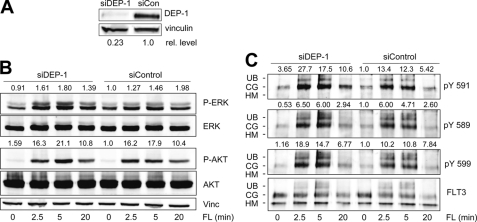
Fms-like tyrosine kinase 3 (FLT3) plays an important role in hematopoietic differentiation, and constitutively active FLT3 mutant proteins contribute to the development of acute myeloid leukemia. Little is known about the protein-tyrosine phosphatases (PTP) affecting the signaling activity of FLT3. To identify such PTP, myeloid cells expressing wild type FLT3 were infected with a panel of lentiviral pseudotypes carrying shRNA expression cassettes targeting different PTP. Out of 20 PTP tested, expressed in hematopoietic cells, or presumed to be involved in oncogenesis or tumor suppression, DEP-1 (PTPRJ) was identified as a PTP negatively regulating FLT3 phosphorylation and signaling. Stable 32D myeloid cell lines with strongly reduced DEP-1 levels showed site-selective hyperphosphorylation of FLT3. In particular, the sites pTyr-589, pTyr-591, and pTyr-842 involved in the FLT3 ligand (FL)-mediated activation of FLT3 were hyperphosphorylated the most. Similarly, acute depletion of DEP-1 in the human AML cell line THP-1 caused elevated FLT3 phosphorylation. Direct interaction of DEP-1 and FLT3 was demonstrated by "substrate trapping" experiments showing association of DEP-1 D1205A or C1239S mutant proteins with FLT3 by co-immunoprecipitation. Moreover, activated FLT3 could be dephosphorylated by recombinant DEP-1 in vitro. Enhanced FLT3 phosphorylation in DEP-1-depleted cells was accompanied by enhanced FLT3-dependent activation of ERK and cell proliferation. Stable overexpression of DEP-1 in 32D cells and transient overexpression with FLT3 in HEK293 cells resulted in reduction of FL-mediated FLT3 signaling activity. Furthermore, FL-stimulated colony formation of 32D cells expressing FLT3 in methylcellulose was induced in response to shRNA-mediated DEP-1 knockdown. This transforming effect of DEP-1 knockdown was consistent with a moderately increased activation of STAT5 upon FL stimulation but did not translate into myeloproliferative disease formation in the 32D-C3H/HeJ mouse model. The data indicate that DEP-1 is negatively regulating FLT3 signaling activity and that its loss may contribute to but is not sufficient for leukemogenic cell transformation.


Figures

FIGURE 1.
FIGURE 1.
Down-regulation of DEP-1 in 32D cells results in enhanced FLT3 signaling and enhanced phosphorylation of FLT3. A, whole cell lysates of 32D cells expressing wild type muFLT3 and DEP-1-specific shRNA (targets A2 or A3) or a nontargeting control shRNA were subjected to SDS-PAGE, blotted to a PVDF membrane, and analyzed with antibodies recognizing DEP-1 and vinculin. Chemiluminescence signals were detected using a CCD camera-based chemiluminescence detection system and calculated relative to vinculin. Relative (rel.) levels of DEP-1 normalized to vinculin are indicated. B, quantitative RT-PCR for DEP-1 mRNA in the above cell lines. Mean ± S.D. of at least three independent experiments. C–F, indicated cell lines were starved for 4 h in serum- and cytokine-free medium and were stimulated with FL (50 ng/ml) for the indicated time periods. C–H, activation of ERK (C and E), AKT (C), STAT5 (D), or PLCγ (G) was analyzed using the indicated phospho-specific antibodies. Blots were reprobed for total signaling proteins. β-Actin was analyzed as loading control. Numbers above the phospho-specific blots represent quantification of the phosphor-specific signals, normalized to the corresponding signals with pan-specific antibodies, and relative to the signal in unstimulated cells harboring control shRNA, which was set to 1.0. The blots shown are representative for at least three experiments with consistent results. F, comparison of phosphorylation of ERK1/2 detected by conventional anti-pThr-202/pTyr-204 antibodies, and anti-pThr-202 antibodies. Ratio between ERK1/2 phosphorylation of shDEP-1 to shControl cells of three independent experiments is shown. H, FLT3 was immunoprecipitated and analyzed by immunoblotting with anti-FLT3, phosphotyrosine (pY100), or phospho-FLT3 (pY591) antibodies. HM, high mannose; CG, complex glycosylated FLT3. Quantification refers to complex glycosylated (surface) FLT3. Representative blots of at least three time repeated experiments are shown.
FIGURE 2.
FIGURE 2.
Down-regulation of DEP-1 causes hyperphosphorylation of human FLT3. 32D cells expressing human FLT3- and DEP-1-specific shRNAs A2 and A3 or a control shRNA were starved and stimulated with FL (50 ng/ml) for indicated time periods. FLT3 was immunoprecipitated and analyzed by immunoblotting with anti-FLT3 or phosphotyrosine (4G-10, A) or phospho-FLT3 (pY591, B) antibodies. HM, high mannose; CG, complex glycosylated FLT3; UB, ubiquitinated. Graphs demonstrate specific phosphorylation of total FLT3 receptor. Phosphospecific to total FLT3 receptor chemiluminescence signals are shown (sum of high mannose, complex glycosylated FLT3, and UB protein). Means ± S.D. are of at least three independent experiments.
FIGURE 3.
FIGURE 3.
Depletion of DEP-1 in THP-1 cells results in elevated FL-mediated signaling activity and FLT3 receptor phosphorylation. Human THP-1 cells were transfected with a siRNA targeting DEP-1 (siDEP-1) or a nontargeting control siRNA (siCon). 48 h post-transfection, cells were starved and stimulated with FL (100 ng/ml) for the indicated time periods. Cells were lysed and subjected to SDS-PAGE and Western blotting. A, cell lysates were probed with antibodies recognizing DEP-1 and vinculin (Vinc.). Depletion of DEP-1 was quantified as ratio of DEP-1 to vinculin. Relative (rel.) levels of DEP-1, normalized to vinculin, are indicated. B, total cell lysates were probed with antibodies recognizing phosphorylated signaling proteins AKT or ERK. Blots were reprobed for total signaling proteins. C, subsequent to stimulation and cell lysis, FLT3 was immunoprecipitated and analyzed by immunoblotting with site-specific phosphotyrosine FLT3 antibodies as indicated and reprobed for total immunoprecipitated FLT3. HM, high mannose; CG, complex glycosylated; UB, ubiquitinated FLT3. Numbers above the phosphotyrosine blots indicate specific phosphorylation of complex glycosylated FLT3. The blots are representative for at least four experiments.
FIGURE 4.
FIGURE 4.
Overexpression of DEP-1 impairs FLT3 signaling and phosphorylation. 32D cells expressing huFLT3, stably transduced with an expression construct for human DEP-1 or the corresponding control vector, were analyzed for DEP-1 expression, FL-mediated signaling activity, and receptor phosphorylation. A, whole cell lysates were blotted with antibodies recognizing both endogenous murine and exogenous human DEP-1 and vinculin for control (Con). B, expression of transduced huDEP-1 was detected by labeling with a monoclonal antibody recognizing a surface epitope of human DEP-1 and subsequent labeling with anti-mouse-Cy3 antibody. Surface-localized DEP-1 was quantified by flow cytometry. C and D, indicated cell lines were starved for 4 h in serum- and cytokine-free medium and were stimulated with FL (50 ng/ml) for the indicated time periods. Whole cell lysates were blotted and activation of ERK or AKT was analyzed using phospho-specific antibodies. Blots were reprobed for total signaling proteins. D, phosphorylation of FLT3 was analyzed by immunoblotting with anti-phosphotyrosine antibody 4G-10 and anti-phospho FLT3 antibody pTyr-591, and blots were subsequently reprobed for total FLT3. HM, high mannose; CG, complex glycosylated FLT3. Representative blots of experiments repeated at least three time are demonstrated.
FIGURE 5.
FIGURE 5.
DEP-1 directly interacts with and dephosphorylates FLT3. A, THP-1 cell were starved in cytokine-free RPMI 1640 medium containing 0.5% FCS, and left unstimulated, or were stimulated with FL (100 ng/ml) for 30 min (FL +). Cells were subsequently lysed, and FLT3 was immunoprecipitated. Immunoprecipitated FLT3 obtained from stimulated cells was incubated with the indicated amounts of recombinant wild type GST-DEP-1 (catalytic domain) fusion protein (WT) or the corresponding catalytically inactive GST-DEP-1 C1239S protein (CS) in the indicated amounts (μg of protein) for 30 min. The samples were subsequently subjected to SDS-PAGE and immunoblotting. Blots were probed with anti-phosphotyrosine antibody (pY100) and reprobed with anti-FLT3 antibody, as indicated. Tyrosine phosphorylation of FLT3 was quantified as the ratio of phosphorylated FLT3 to total FLT3 (numbers above pY100 blot). This experiment was performed twice with consistent results. B, HEK293 cells were co-transfected with expression plasmids for human FLT3 and wild type (WT) DEP-1 or catalytically inactive DEP-1 C1239S (CS) in the indicated amounts (μg) using polyethyleneimine. 36 h after transfection, cells were starved for 8 h and then stimulated with 100 ng/ml FL for 5 min. The cells were subsequently lysed, and FLT3 was immunoprecipitated (IP). The blots were probed for phosphorylation of FLT3 by immunoblotting and reblotted for the amounts of FLT3. Quantification denotes the FLT3 phosphorylation signal normalized to the amounts of FLT3, relative to the control in absence of DEP-1. Co-purified DEP-1 protein in the immunoprecipitates was analyzed using anti-DEP-1 antibody. DEP-1 expression in the cell lysates was also analyzed (lowest panel). HM, high mannose; CG, complex glycosylated FLT3. A representative experiment of three with consistent results is depicted. C, HEK293 cells were co-transfected with plasmids encoding human FLT3 along with wild type DEP-1 (WT) or the DEP-1 D1205A trapping mutant (DA, as indicated) in the molar ratio 1:1. The cells were starved and stimulated with FL as in A and lysed, and either DEP-1 (upper panel) or FLT3 (lower panel) was immunoprecipitated. For a specificity control, immunoprecipitation was carried out with unspecific IgG. Immunoprecipitated and co-immunoprecipitated proteins were analyzed by immunoblotting with anti-FLT3, anti-DEP-1, or anti-phosphotyrosine (pY100) antibodies as indicated. HM, high mannose; CG, complex glycosylated FLT3. A representative experiment of three with consistent results is shown.
FIGURE 6.
FIGURE 6.
Depletion of DEP-1 stimulates proliferation and clonal growth of 32D cell lines. A, 32D cells expressing wild type muFLT3 and DEP-1 shRNA A2 or control shRNA were seeded in 96-well plates and cell growth in the absence of cytokines, in the presence of FL (20 ng/ml), or in the presence of IL-3 (2 ng/ml) and was measured after 2 days using the MTT method. B, cells were seeded in 96-well plates at a density of 0.8 × 10⁵ per ml in RPMI 1640 medium supplemented with 10% fetal calf serum without cytokines, with FL (20 ng/ml), or IL-3 (2 ng/ml) as indicated. Cell growth was measured at the indicated time points by measuring cellular GFP fluorescence. Values are means of triplicates, and the shown data set is representative for three experiments with consistent results. C, clonal growth of 32D cells in methylcellulose. The above cell pools were subjected to colony formation assays in methylcellulose, in cytokine in the absence or presence of FL (20 ng/ml) or IL-3 (2.5 ng/ml). Representative sections were photographed after 12 days (FL samples) or 6 days (IL-3 samples) of culture, and colonies were quantified by counting representative fields of the wells.

References

    1. Stirewalt D. L., Radich J. P. (2003) Nat. Rev. Cancer 3, 650–665 - PubMed
    1. Schmidt-Arras D., Schwäble J., Böhmer F. D., Serve H. (2004) Curr. Pharm. Des. 10, 1867–1883 - PubMed
    1. Gilliland D. G., Griffin J. D. (2002) Blood 100, 1532–1542 - PubMed
    1. Thiede C., Steudel C., Mohr B., Schaich M., Schäkel U., Platzbecker U., Wermke M., Bornhäuser M., Ritter M., Neubauer A., Ehninger G., Illmer T. (2002) Blood 99, 4326–4335 - PubMed
    1. Small D. (2006) Hematology Am. Soc. Hematol. Educ. Program, 178–184 - PubMed

Publication types

MeSH terms

Substances

LinkOut - more resources