. 2011 Apr 8;286(14):12123-32.
doi: 10.1074/jbc.M110.154625. Epub 2011 Jan 24.

Erythropoietin protects intestinal epithelial barrier function and lowers the incidence of experimental neonatal necrotizing enterocolitis

Affiliations

Erythropoietin protects intestinal epithelial barrier function and lowers the incidence of experimental neonatal necrotizing enterocolitis

Sheng-Ru Shiou et al. J Biol Chem. .

Abstract

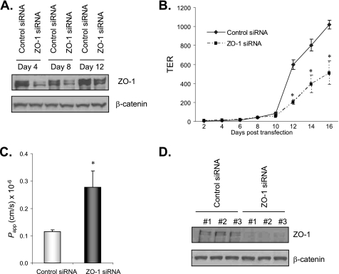
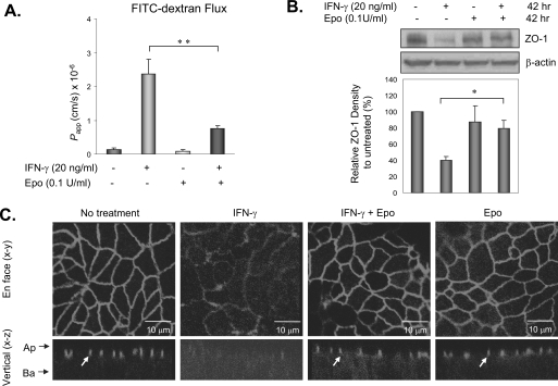
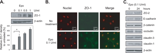
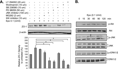
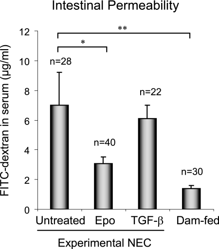
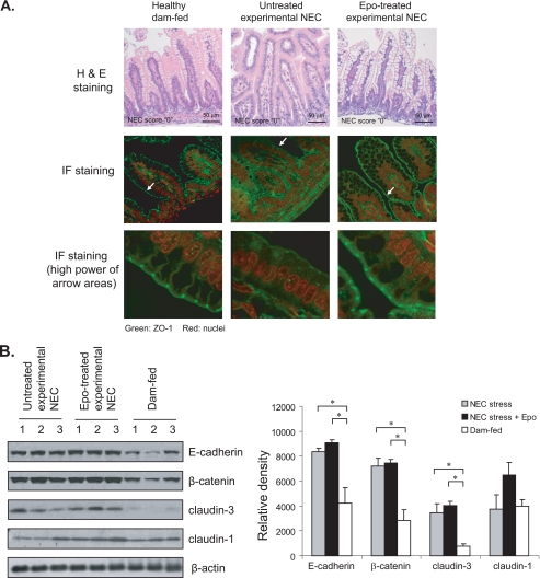
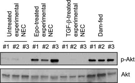
The impermeant nature of the intestinal barrier is maintained by tight junctions (TJs) formed between adjacent intestinal epithelial cells. Disruption of TJs and loss of barrier function are associated with a number of gastrointestinal diseases, including neonatal necrotizing enterocolitis (NEC), the leading cause of death from gastrointestinal diseases in preterm infants. Human milk is protective against NEC, and the human milk factor erythropoietin (Epo) has been shown to protect endothelial cell-cell and blood-brain barriers. We hypothesized that Epo may also protect intestinal epithelial barriers, thereby lowering the incidence of NEC. Our data demonstrate that Epo protects enterocyte barrier function by supporting expression of the TJ protein ZO-1. As immaturity is a key factor in NEC, Epo regulation of ZO-1 in the human fetal immature H4 intestinal epithelial cell line was examined and demonstrated Epo-stimulated ZO-1 expression in a dose-dependent manner through the PI3K/Akt pathway. In a rat NEC model, oral administration of Epo lowered the incidence of NEC from 45 to 23% with statistical significance. In addition, Epo treatment protected intestinal barrier function and prevented loss of ZO-1 at the TJs in vivo. These effects were associated with elevated Akt phosphorylation in the intestine. This study reveals a novel role of Epo in the regulation of intestinal epithelial TJs and barrier function and suggests the possible use of enteral Epo as a therapeutic agent for gut diseases.


Figures

FIGURE 1.
FIGURE 1.
ZO-1 silencing impaired TER and barrier function in enterocytes. A, ZO-1 siRNA transfection decreased ZO-1 protein expression. T84 cells at 50% confluence were transfected with 200 nm of a pool of four ZO-1 or scrambled control siRNAs. Cell lysates were collected on days 4, 8, and 12 after transfection for immunoblotting for ZO-1 and β-catenin. B, ZO-1 silencing decreased TER. Cells were plated in Transwells in triplicate along with transfection reagents and 200 nm ZO-1 or control siRNAs. Medium was changed the next day and then twice a week. TER was monitored over time using a voltohmmeter. C, ZO-1 silencing impaired barrier function. Cells were grown and transfected as in B and barrier function was determined as described under “Materials and Methods” on day 16 when control siRNA-transfected cells reached high TER (>1,000 ohms·cm2). Cell lysates from each Transwell were then collected for ZO-1 immunoblotting in D. Representative immunoblots from three experiments with similar results are shown. Data from three experiments are presented as mean ± S.E., and * depicts statistical significance with a p value < 0.05 by t test.
FIGURE 2.
FIGURE 2.
Epo protected enterocyte barrier function and ZO-1 localization from inflammatory cytokine-induced damage. A, Epo attenuated IFN-γ-induced barrier disruption. T84 human enterocytes were plated in Transwells in triplicate to reach a high TER and treated with 20 ng/ml IFN-γ ± 0.1 unit/ml Epo for 72 h. Barrier function was then determined by apical to basolateral flux of 10-kDa FITC-dextran, and apparent permeability coefficients (Papp) were calculated as described under “Materials and Methods.” B, Epo rescued IFN-γ down-regulation of ZO-1. T84 cells were grown and treated as in A for 42 h and lysed. The lysates were used for immunoblotting for ZO-1 and β-actin. C, confocal microscopy of ZO-1 localization in T84 cells with or without 100 ng/ml IFN-γ and 0.1 unit/ml Epo treatments for 72 h. Images shown are the en face (x-y) and vertical (x-z) sections. TJs shown in the vertical sections are designated by arrows. Arrows by the x-z sections indicate the apical (Ap) and basal (Ba) aspects of the cells. Representative immunoblots from three experiments with similar results are shown. Data from three experiments are presented as mean ± S.E. relative to controls in percentage. * and ** depict statistical significance with p values < 0.05 and <0.01, respectively, by one-way ANOVA with Bonferroni correction.
FIGURE 3.
FIGURE 3.
Epo increased TJ protein ZO-1 expression in human fetal immature IEC. A and C, H4 immature IEC were grown on tissue culture plates and treated with 0, 0.1, 0.5, and 1 unit/ml recombinant human Epo for 24 h. Total cell lysates were collected and subjected to immunoblotting for ZO-1 and β-actin in A and ZO-2, E-cadherin, β-catenin, occludin, claudin-1, claudin-3, and β-actin in C. B, H4 cells grown on chamber slides were treated with or without 0.1 unit/ml Epo for 24 h and then subjected to immunofluorescence staining for ZO-1 and nuclei. Images were acquired by confocal microscopy. Representative immunoblots from three experiments with similar results are shown. Data from three experiments are presented as mean ± S.E. relative to controls in percentage. * depicts statistical significance with a p value <0.05 by one-way ANOVA with Bonferroni correction..
FIGURE 4.
FIGURE 4.
Epo stimulated ZO-1 expression via the PI3K/Akt pathway in human fetal immature IEC. A, effect of inhibitors on Epo regulation of ZO-1 expression in immature IEC. H4 cells were left untreated or pretreated with inhibitors as indicated for 30 min and then together with 0.1 unit/ml Epo for another 24 h. Cell lysates were collected and subjected to immunoblotting for ZO-1 and β-actin. B, Epo activated the Akt pathway in immature IEC. H4 cells were treated with 0.1 unit/ml Epo for various periods of time as indicated and then lysed. Lysates were used for immunoblotting for p-Akt, total Akt, p-JNK, total JNK, p-ERK1/2, and total ERK1/2. Representative immunoblots from three experiments with similar results are shown. Data from three experiments are presented as mean ± S.E. relative to controls in percentage. * depicts statistical significance with a p value <0.05, by one-way ANOVA with Bonferroni correction
FIGURE 5.
FIGURE 5.
Epo protected intestinal barrier function in an NEC animal model. Preterm neonatal rats delivered by cesarean section from Sprague-Dawley dams were fed with formula ± 0.1 unit/ml Epo or 30 ng/ml TGF-β (as a treatment control for Epo) and stressed to induce NEC. Naturally born, unstressed, and dam-fed pups were included as healthy controls. In vivo barrier function was determined in all surviving pups at the end of the animal experiment on day 5 as described under “Materials and Methods.” Data from seven animal experiments are presented as mean ± S.E. * and **depict statistical significance with p values <0.05 and 0.01, respectively, by one-way ANOVA with Bonferroni correction. Higher levels of the tracer FITC-dextran in blood indicate poorer intestinal barrier function.
FIGURE 6.
FIGURE 6.
Effect of Epo on the localization and expression of adherens and tight junction proteins in a NEC animal model. A, Epo maintained ZO-1 at the intestinal tight junction. Intestinal sections were collected from animals used for in vivo barrier function as in Fig. 5 and subjected to H&E staining and immunofluorescence (IF) staining for ZO-1 and nuclei. Images were acquired by confocal microscopy. Representative immunofluorescence staining shows expression and localization of ZO-1 at the TJs of healthy and Epo-treated experimental NEC intestines but not in the non-Epo-treated NEC stressed samples. H&E staining shows NEC score 0 for all sections. B, Epo did not alter the expression of E-cadherin, β-catenin, claudin-1, and claudin-3 in experimental NEC animals. Intestinal lysates were collected from animals used for in vivo barrier function as in Fig. 5 and subjected to immunoblotting for E-cadherin, β-catenin, claudin-1, claudin-3, and β-actin. Representative immunoblots from three experiments with similar results are shown. * depicts statistical significance with a p value < 0.05, by one-way ANOVA with Bonferroni correction.
FIGURE 7.
FIGURE 7.
Epo activated the Akt pathway in vivo in the NEC animal model. Intestinal lysates were collected from animals used for in vivo barrier function as in Fig. 5 and subjected to immunoblotting for phospho- and total Akt. Representative immunoblots from three experiments with similar results are shown.

References

    1. Kosloske A. M. (1994) Acta Paediatr. Suppl. 396, 2–7 - PMC - PubMed
    1. Holman R. C., Stehr-Green J. K., Zelasky M. T. (1989) Am. J. Public Health 79, 987–989 - PMC - PubMed
    1. Anand R. J., Leaphart C. L., Mollen K. P., Hackam D. J. (2007) Shock 27, 124–133 - PubMed
    1. Anderson J. M., Balda M. S., Fanning A. S. (1993) Curr. Opin. Cell Biol. 5, 772–778 - PubMed
    1. Stevenson B. R., Siliciano J. D., Mooseker M. S., Goodenough D. A. (1986) J. Cell Biol. 103, 755–766 - PMC - PubMed

Publication types

MeSH terms