Case Reports
. 2011 Jan 25;76(4):327-36.
doi: 10.1212/WNL.0b013e31820882bd.

Myasthenic syndrome caused by plectinopathy

Affiliations
Case Reports

Myasthenic syndrome caused by plectinopathy

D Selcen et al. Neurology. .

Abstract

Background: Plectin crosslinks intermediate filaments to their targets in different tissues. Defects in plectin cause epidermolysis bullosa simplex (EBS), muscular dystrophy (MD), and sometimes pyloric atresia. Association of EBS with a myasthenic syndrome (MyS) was documented in a single patient in 1999.

Objectives: To analyze the clinical, structural, and genetic aspects of a second and fatal case of EBS associated with a MyS and search for the genetic basis of the disease in a previously reported patient with EBS-MD-MyS.

Methods: Clinical observations; histochemical, immunocytochemical, and electron microscopy studies of skeletal muscle and neuromuscular junction; and mutation analysis.

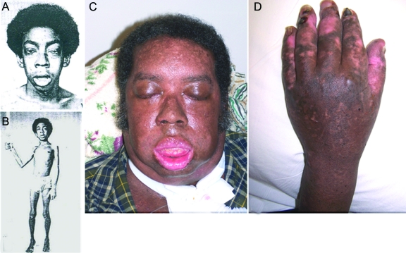
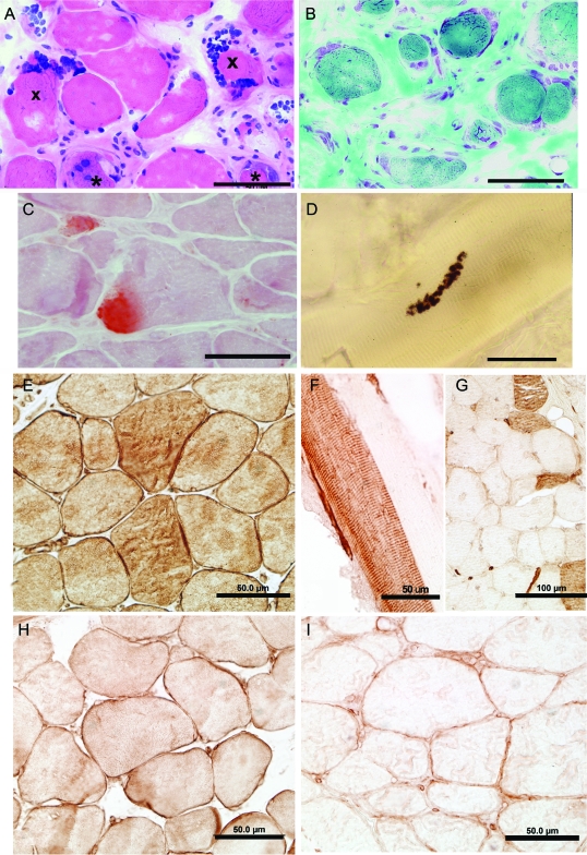
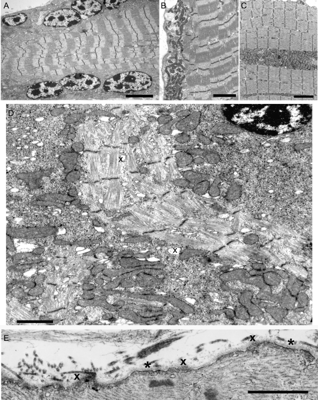
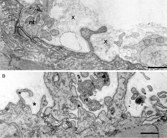
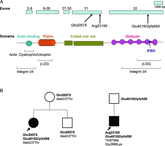
Results: An African American man had EBS since early infancy, and progressive muscle weakness, hyperCKemia, and myasthenic symptoms refractory to therapy since age 3 years. Eventually he became motionless and died at age 42 years. At age 15 years, he had a marked EMG decrement, and a reduced miniature endplate potential amplitude. The myopathy was associated with dislocated muscle fiber organelles, structurally abnormal nuclei, focal plasmalemmal defects, and focal calcium ingress into muscle fibers. The neuromuscular junctions showed destruction of the junctional folds, and remodeling. Mutation analysis demonstrated a known p.Arg2319X and a novel c.12043dupG mutation in PLEC1. The EBS-MD-MyS patient reported in 1999 also carried c.12043dupG and a novel p.Gln2057X mutation. The novel mutations were absent in 200 Caucasian and 100 African American subjects.

Conclusions: The MyS in plectinopathy is attributed to destruction of the junctional folds and the myopathy to defective anchoring of muscle fiber organelles and defects in sarcolemmal integrity.


Figures

Figure 1
Figure 1. Patient photographs
(A, B) Patient at age 17 years. Note severe asymmetric bilateral ptosis, hyperactive frontalis muscle, facial paresis, open mouth, cubitus valgus, Achilles tendon contractures, and diffuse muscle atrophy. (C, D) Patent at age 41 years. He has a tracheostomy, has facial diplegia, is unable to close his mouth or open his eyes, and shows the chronic skin changes of epidermolysis bullosa simplex. He also has blisters on his lip and tongue and oral moniliasis.
Figure 2
Figure 2. Histochemistry and plectin localization studies in patient 2
(A, B) Note marked variation in fiber size, regenerating fiber elements (asterisks), endomysial fibrosis (B), and clusters of large nuclei at periphery of several fibers. (C) Alizarin red stain reveals focal calcium deposits in 2 fibers. (D) Multiple small cholinesterase-reactive endplate regions arrayed over an extended length of the fiber. Plectin was localized in normal control muscle (E, H) and patient intercostal muscle (F, G, I) with antibody recognizing the plectin rod domain (anti-Rod Ab) (E–G) and antibody recognizing the C-terminal plectin domain (anti-C Ab) (H, I). (E, H) In normal muscle, plectin is localized to the sarcolemma and sarcoplasm with both Abs. The anti-Rod Ab shows plectin-depleted and plectin-positive muscle fibers (F, G), whereas the anti-C Ab shows sarcoplasmic loss and slight sarcolemmal expression of plectin in all muscle fibers (I). Bars indicate 50 μm in all panels except in (G), where they indicate 100 μm.
Figure 3
Figure 3. Ultrastructural findings in abnormal muscle fibers of patient 2
(A) Note subsarcolemmal rows of large nuclei harboring multiple prominent chromatin bodies. (B, C) Subsarcolemmal and intrafiber clusters of mitochondria surrounded by fiber regions devoid of mitochondria. (D) Aberrant and disrupted myofibrils surrounded by clusters of mitochondria intermingled with glycogen, ribosomes, and dilated vesicles (x). Note preapoptotic nucleus at upper right. (E) Focal sarcolemma defects due to gaps in the plasma membrane. Where the plasma membrane is absent, the overlying basal lamina is thickened (x). Small vesicles underlie the thickened basal lamina. Asterisks indicate segments of the preserved plasma membrane. Bars = 4 μm in (A), 3 μm (B, C), 1.4 μm in (D), 1 μm in (E).
Figure 4
Figure 4. Abnormal endplate (EP) regions in patient 2
(A) The imaged EP regions show partial occupancy of the postsynaptic region by the nerve terminal and remnants of degenerate folds (x). The nerve terminal (nt) occupies only part of the postsynaptic region. Degenerate remnants of the folds (x) appear over the simplified postsynaptic region from which folds were lost. Dark reaction product on postsynaptic membrane shows acetylcholine receptor localization with peroxidase labeled α-bungarotoxin. (B) Small nerve terminal occupies only part of a highly simplified postsynaptic region. Asterisk indicates remnants of basal lamina that surrounded preexisting folds. Bars = 1 μm.
Figure 5
Figure 5. Plectin domains and identified PLEC variants
(A) Schematic representation of PLEC exons 2–32 indicating identified patient mutations and binding domains associated with C- and N-terminal regions of plectin. (B) Family analysis of P1 and P2 shows transmission of pathogenic mutations (bold face) and polymorphisms.

References

    1. Elliott CE, Becker B, Oehler S, Castanon MJ, Hauptmann R, Wiche G. Plectin transcript diversity: identification and tissue distribution of variants with distinct first coding exons and rodless isoforms. Genomics 1997;42:115–125 - PubMed
    1. Fuchs P, Zorer M, Rezniczek GA, et al. Unusual 5′ transcript complexity of plectin isoforms: novel tissue-specific exons modulate actin binding activity. Hum Mol Genet 1999;8:2461–2472 - PubMed
    1. Konieczny P, Fuchs P, Reipert S, et al. Myofiber integrity depends on desmin network targeting to Z-disks and costameres via distinct plectin isoforms. J Cell Biol 2008;181:667–681 - PMC - PubMed
    1. Banwell BL, Russel J, Fukudome T, Shen X-M, Stilling G, Engel AG. Myopathy, myasthenic syndrome, and epidermolysis bullosa simplex due to plectin deficiency. J Neuropathol Exp Neurol 1999;58:832–846 - PubMed
    1. McLean W, Pulkkinen L, Smith F, et al. Loss of plectin causes epidermolysis bullosa with muscular dystrophy: cDNA cloning and genomic organization. Genes Dev 1996;10:1724–1735 - PubMed

Publication types

MeSH terms