

Computer-aided detection of early interstitial lung diseases using low-dose CT images
- PMID: 21263171
- PMCID: PMC3165025
- DOI: 10.1088/0031-9155/56/4/016
Computer-aided detection of early interstitial lung diseases using low-dose CT images
Abstract
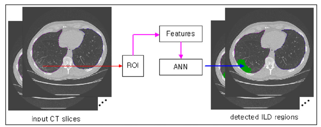
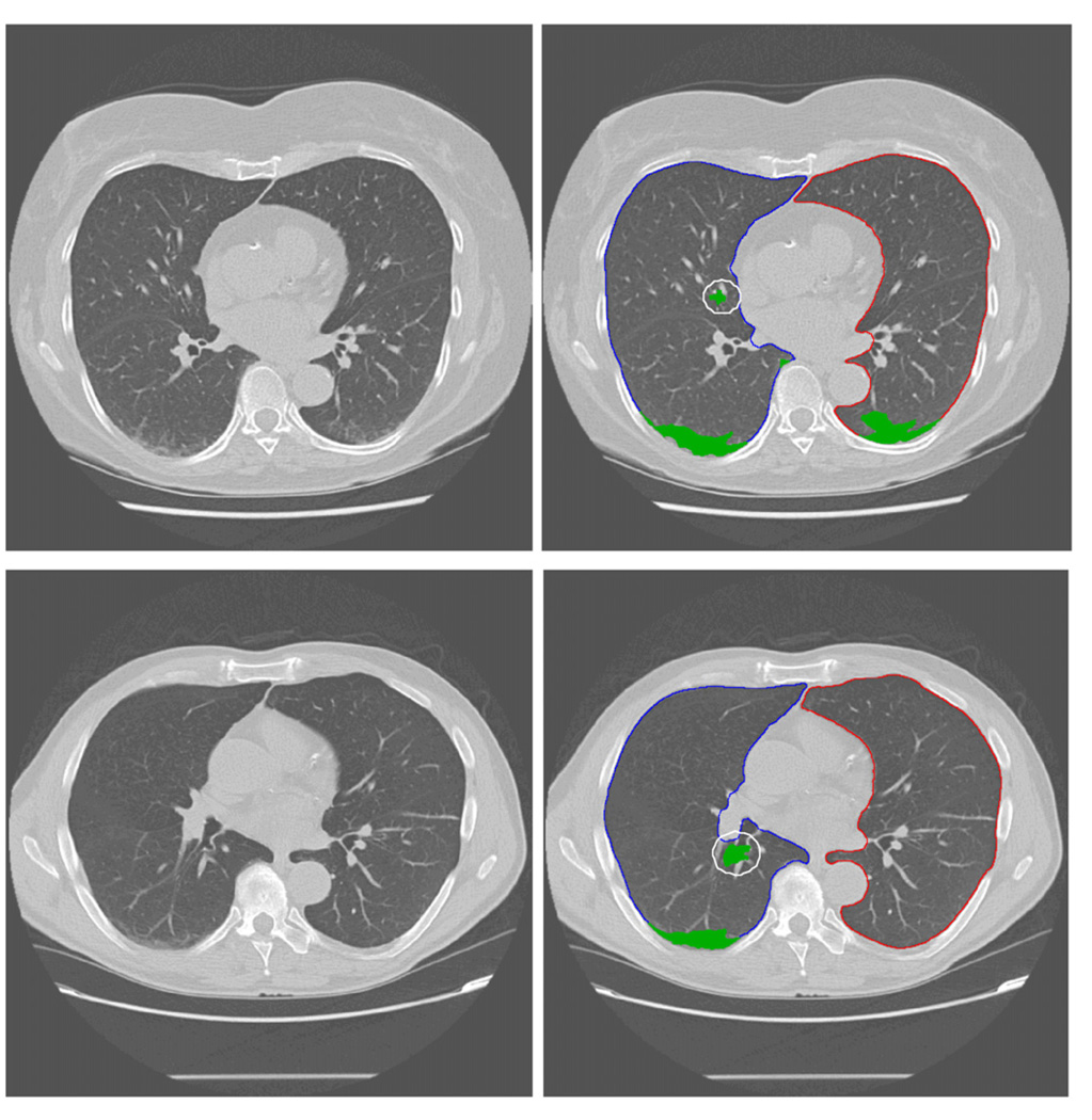
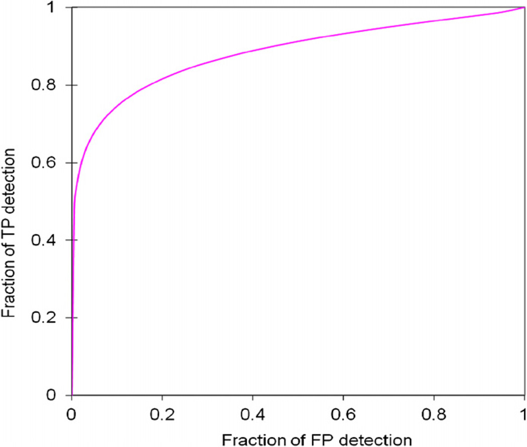
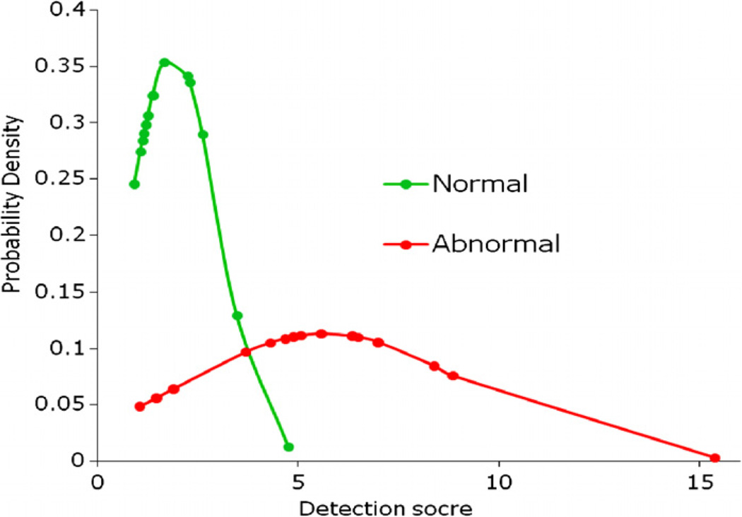
This study aims to develop a new computer-aided detection (CAD) scheme to detect early interstitial lung disease (ILD) using low-dose computed tomography (CT) examinations. The CAD scheme classifies each pixel depicted on the segmented lung areas into positive or negative groups for ILD using a mesh-grid-based region growth method and a multi-feature-based artificial neural network (ANN). A genetic algorithm was applied to select optimal image features and the ANN structure. In testing each CT examination, only pixels selected by the mesh-grid region growth method were analyzed and classified by the ANN to improve computational efficiency. All unselected pixels were classified as negative for ILD. After classifying all pixels into the positive and negative groups, CAD computed a detection score based on the ratio of the number of positive pixels to all pixels in the segmented lung areas, which indicates the likelihood of the test case being positive for ILD. When applying to an independent testing dataset of 15 positive and 15 negative cases, the CAD scheme yielded the area under receiver operating characteristic curve (AUC = 0.884 ± 0.064) and 80.0% sensitivity at 85.7% specificity. The results demonstrated the feasibility of applying the CAD scheme to automatically detect early ILD using low-dose CT examinations.
Figures
References
-
- Arzhaeva Y, et al. Computer-aided detection of interstitial abnormalities in chest radiographs using a reference standard based on computed tomography. Med. Phys. 2007;34:4798–4809. - PubMed
-
- Aziz ZA, et al. Functional impairment in emphysema: contribution of airway abnormalities and distribution of parenchymal disease. Am. J. Roentgenol. 2005;185:1509–1515. - PubMed
-
- Best AC, et al. Quantitative CT indexes in idiopathic pulmonary fibrosis: relationship with physiologic impairment. Radiology. 2003;228:407–414. - PubMed
-
- Biederer J, et al. Correlation between HRCT finding, pulmonary function tests and bronchoalveolar lavage cytology in interstitial lung disease associated with rheumatoid arthritis. Eur. Radiol. 2004;14:272–280. - PubMed
-
- Bjoraker JA, et al. Prognostic significance of histopathologic subsets in idiopathic pulmonary fibrosis. Am. J. Respir. Crit. Care Med. 1998;157:199–203. - PubMed
Publication types
MeSH terms
Grants and funding
LinkOut - more resources
Full Text Sources
Medical
Miscellaneous