Review
. 2011 Mar;21(3):421-34.
doi: 10.1038/cr.2011.14. Epub 2011 Jan 25.

The double face of the histone variant H3.3

Affiliations
Review

The double face of the histone variant H3.3

Emmanuelle Szenker et al. Cell Res. 2011 Mar.

Abstract

Histone proteins wrap DNA to form nucleosome particles that compact eukaryotic genomes while still allowing access for cellular processes such as transcription, replication and DNA repair. Histones exist as different variants that have evolved crucial roles in specialized functions in addition to their fundamental role in packaging DNA. H3.3--a conserved histone variant that is structurally very close to the canonical histone H3--has been associated with active transcription. Furthermore, its role in histone replacement at active genes and promoters is highly conserved and has been proposed to participate in the epigenetic transmission of active chromatin states. Unexpectedly, recent data have revealed accumulation of this specific variant at silent loci in pericentric heterochromatin and telomeres, raising questions concerning the actual function of H3.3. In this review, we describe the known properties of H3.3 and the current view concerning its incorporation modes involving particular histone chaperones. Finally, we discuss the functional significance of the use of this H3 variant, in particular during germline formation and early development in different species.


Figures
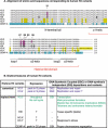
Figure 1
Figure 1
Sequence alignment and specific features of human H3 variants. (A) Alignment of amino acid sequences corresponding to human H3 variants. Sequences are compared with the “ancestral” variant H3.3 and the amino acid differences are highlighted. H3.1 and H3.2 differences are highlighted in purple, H3t in gray, H3.X and H3.Y in yellow, and CENP-A in light blue. The position numbers of amino acids that are different between H3.3 and H3.1/2 are indicated. The positions of the N-terminal tail and of the α-helixes of the histone-fold motif are shown. (B) Distinct features of human H3 variants. The features of canonical and replacement H3 variants are indicated according to their expression, mode of deposition and contexts. Canonical histones are shown in purple while H3.3 in green.
Figure 2
Figure 2
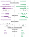
Human histone H3.3 compared with H3.1 and H3.2. Differences between the canonical H3 variants (H3.1 and H3.2) ndash; in purple – and the replacement variant H3.3 – in green – are illustrated. Canonical histone genes are organized in tandem and the cluster HIST1 located on chromosome 6p21 contains 6 histone H1 and 49 core histone genes including 10 histone H3 genes. Canonical histone genes lack introns and are not polyadenylated in contrast to the regular genes coding for H3.3 (H3.3A and H3.3B). The amino acid differences between the canonical H3 and H3.3 are illustrated. H3.3 S31 can be phosphorylated. The motifs SVM and AIG in H3.1/2 and H3.3, respectively, could account for chaperone specificity. We also illustrate the distinct enriched marks in H3 and H3.3 before and after deposition into chromatin.
Figure 3
Figure 3
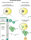
Local enrichment of H3.3 and complexes promoting deposition. Left: in mouse somatic and embryonic cells, H3.3 is enriched in coding regions and at specific chromatin landmarks. In heterochromatin, DAXX cooperates with the chromatin remodeler ATRX in accumulating H3.3 at pericentric heterochromatin and telomeres. It has to be noticed that accumulation of H3.3 at telomeres is so far only described in ES cells. In euchromatin, the HIRA complex mediates H3.3 enrichment in the body of transcribed genes and at promoters of transcribed or non-transcribed genes. The chaperone complex that mediates H3.3 enrichment at regulatory elements remains to be clearly identified but DAXX and/or DEK proteins have been suggested to play a role in this process. Right: in gametes, H3.3 is enriched in sex chromosomes during mouse male meiosis during MSCI. HIRA and DAXX colocalize with XY bodies but their potential role in this process still needs to be uncovered. In zygotes, H3.3 is loaded in the male pronucleus at fertilization in Drosophila and in mouse through the HIRA complex that cooperates with the chromatin remodeling factor CHD1.
Figure 4
Figure 4
Emergence of non-centromeric H3 variants and specialization of their functions during evolution. Schematic representation of the emergence of new H3 histone variants concomitant with the specialization of H3.3 functions through evolution. Most probably, all non-centromeric H3 derive from the ancestral H3.3-like histone (green), whose functions get specialized when new H3 variants emerged, in particular the canonical H3.1 and H3.2 histones (purple). The specialization of canonical H3 and H3.3 variants functions from the universal H3.3-like histone is illustrated.

References

    1. Kornberg RD. Chromatin structure: a repeating unit of histones and DNA. Science. 1974;184:868–871. - PubMed
    1. Luger K, Mader AW, Richmond RK, Sargent DF, Richmond TJ. Crystal structure of the nucleosome core particle at 2.8 A resolution. Nature. 1997;389:251–260. - PubMed
    1. Kouzarides T. Chromatin modifications and their function. Cell. 2007;128:693–705. - PubMed
    1. Zweidler A.Core histone variants of the mouse: primary structure and differential expressionIn: JL Stein GS Stein, WF Marzluff, eds. Vol. Chapter 14, Histones Genes, Structure, Organization and Regulation. John Wiley & sons, 1984339–371.
    1. Talbert PB, Henikoff S. Histone variants - ancient wrap artists of the epigenome. Nat Rev Mol Cell Biol. 2010;11:264–275. - PubMed

Publication types