

Cadmium induces changes on ACTH and PRL cells in Podarcis sicula lizard pituitary gland
- PMID: 21263744
- PMCID: PMC3167330
- DOI: 10.4081/ejh.2010.e45
Cadmium induces changes on ACTH and PRL cells in Podarcis sicula lizard pituitary gland
Abstract
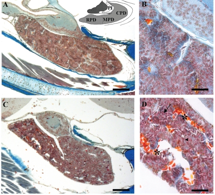
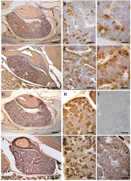
We analyzed the effect of cadmium on corticotropic (ACTH) and prolactin (PRL) cells in the pituitary gland of the Podarcis sicula lizard under chronic exposure to this metal. Adult lizards were given CdCl2 in drinking water at the dose of 10 μg/10 g body mass for 120 days. Light microscopy was performed after histological and immunohistochemical staining, and the effects were followed at regular time intervals up to 120 days post-treatment. We detected substantial variations in the general morphology of the pituitary: unlike the control lizards in which the gland appeared compact, the treated lizards showed a glandular tissue with dilated spaces that were more extensive at 90 and 120 days. PRL and ACTH cells showed an increase in occurrence and immunostaining intensity in treated lizards in comparison with the same cells of control animals. This cellular increase peaked for PRL at 30 days in the rostral, medial and also caudal pars distalis of the gland. ACTH cells appeared to increase markedly after 60 days of treatment in both the pars distalis and the pars intermedia. Again, at 60 days small, isolated ACTH cells were also found in the caudal pars distalis in which these cells were generally absent. However, at 120 days both these cellular types showed an occurrence, distribution and morphology similar to those observed in the control lizards. In lizards, protracted oral exposure to cadmium evidently involves an alteration of the normal morphology of the gland and an inhibitory effect of ACTH and PRL cells, since they increase in occurrence and immunostaining. Yet in time the inhibitory effect of cadmium on ACTH and PRL cells falls back and their occurrence appears similar to that of the control lizard.
Figures
References
-
- Burger J, Campbell KR, Murray S, Campbell TS, Gaines KF, Jeitner C, et al. Metal levels in blood, muscle and liver of water snakes (Nerodia spp.) from New Jersey, Tennessee and South Carolina. Sci Total Environ. 2007;373:556–63. - PubMed
-
- Brzoska MM, Moniuszko-Jakoniuk J. Bone metabolism of male rats chronically exposed to cadmium. Toxicol Appl Pharmacol. 2005;207:195–211. - PubMed
-
- Uriu K, Kaizu K, Komine N, Ikeda M, Qie YL, Hashimoto O, et al. Renal hemodynamics in rats with cadmium-induced nephropathy. Toxicol Appl Pharmacol. 1998;150:76–85. - PubMed
-
- Antonio MT, Corpas I, Leret ML. Neurochemical changes in newborn rat’s brain after gestational cadmium and lead exposure. Toxicol Lett. 1999;104:1–9. - PubMed
MeSH terms
Substances
LinkOut - more resources
Full Text Sources
Research Materials
Miscellaneous
