

NADPH-diaphorase expression in the meibomian glands of rat palpebra in postnatal development
- PMID: 21263746
- PMCID: PMC3167320
- DOI: 10.4081/ejh.2010.e47
NADPH-diaphorase expression in the meibomian glands of rat palpebra in postnatal development
Abstract
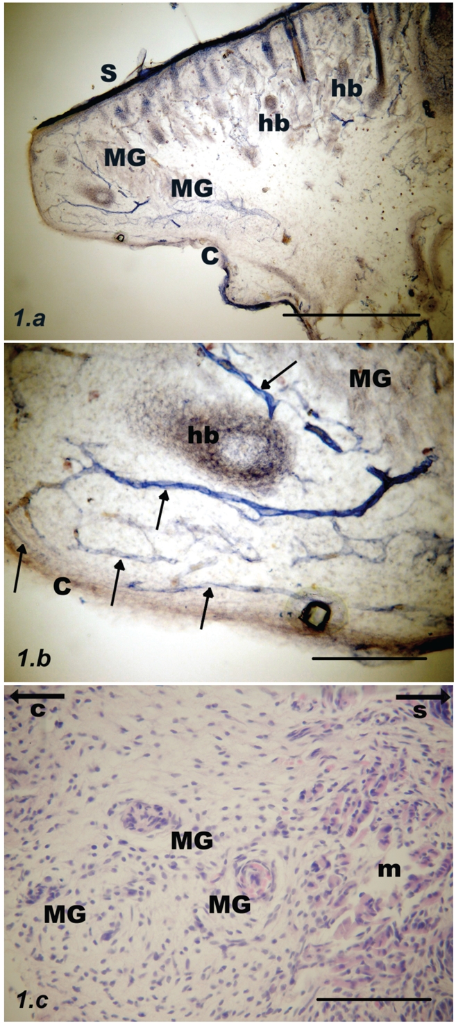
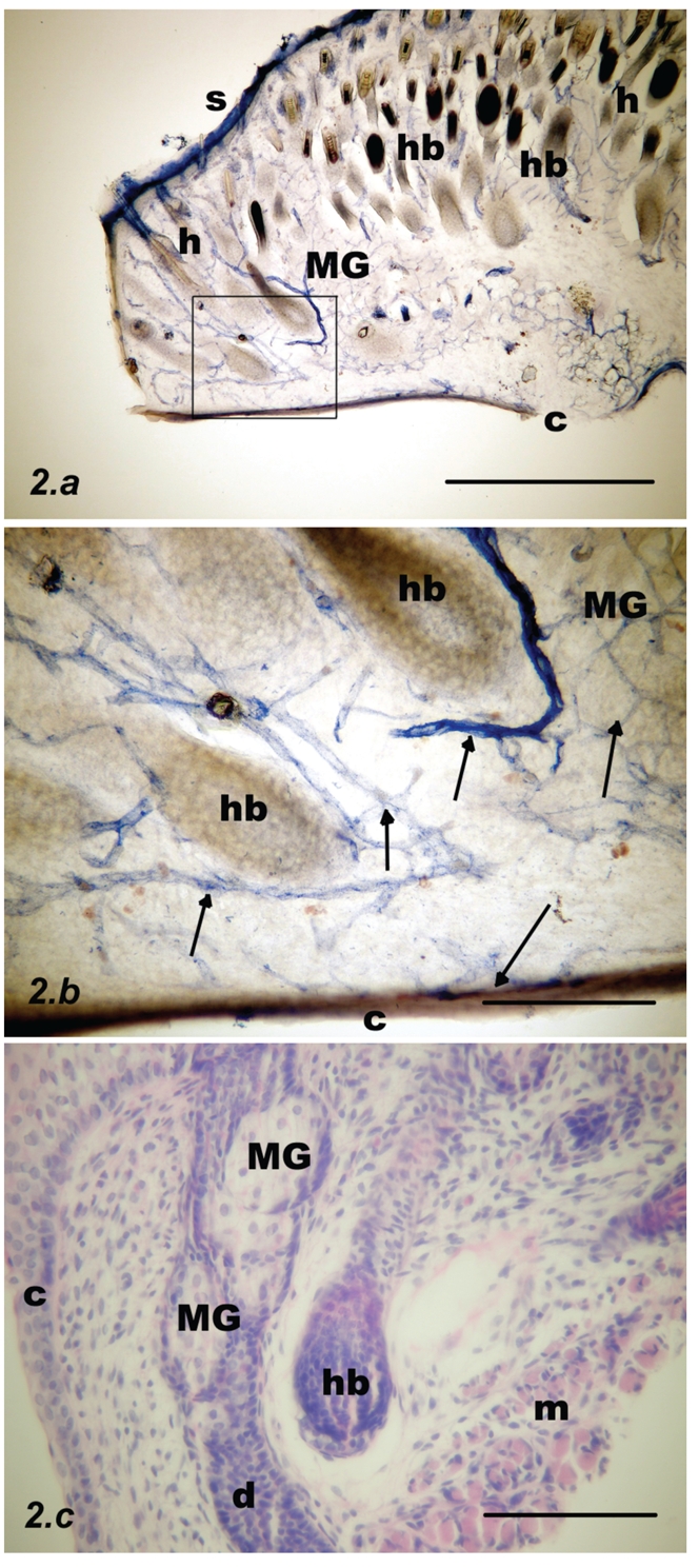
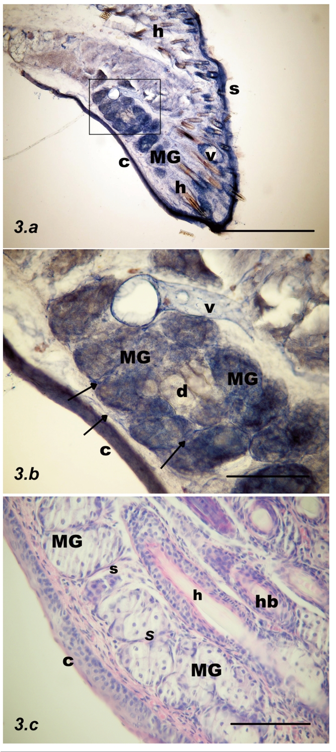
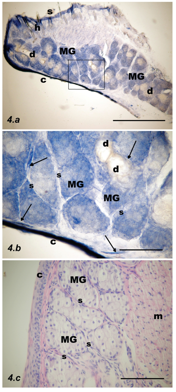
In the current study, we aimed at investigating the presence of nitric oxide synthase (NOS) positive nerve fibers in rat meibomian glands (MGs) at various stages of development. There is good evidence to suggest that nicotinamide adenine dinucleotide phosphate diaphorase (NADPH-d) is a surrogate for neuronal nitric oxide synthase (NOS). Sections of the central, upper eyelids of Wistar rats were processed histochemically for NADPH-d to investigate the presence and distribution of NOS-positive nerve fibers at the following time points: day 1 and weeks 1, 2 and 3 post partum, and in adult controls. At day 1, MG acini were lightly stained and located at a distance from the mucosal border. Vessels were accompanied by intensely stained NADPH-d positive nerve fibers. At the week 1 time point, both the vessels and the NADPH-d positive fibers were still present, but less numerous. MGs were now closer to the mucosa, so that the submucosa was thinner. The acini were mostly pale but occasionally darker. At week 3, there were fewer blood vessels in both the submucosa and within the septa. Darker acini were more common than lightly stained acini. NADPH-d positive dots were observed in the vicinity of the MGs. At the week 3 time point, MGs were adjacent to the mucosal border and stained more intensely than at earlier times; almost all acini were stained. The microscopic appearances were almost identical with those of adult palpebra. Submucosal and septal blood vessels and NADPH-d positive nerve fibers were less numerous. NADPH-d histochemical staining confirmed differences in the density of stained nerve fibers at different developmental stages. The greatest density of NADPH-d -positive nerve fibers occurred in 1-day-old rats whereas they were less numerous in adult rat eyelids. Nerves innervating MGs utilize nitric oxide (NO) as a neurotransmitter mostly in early developmental stages and this need thereafter decreases and stabilizes at 3 weeks postnatally.
Figures
References
-
- Bron A, Tripathi R, Tripathi B. Wolff's Anatomy of the Eye and Orbit. Chapman & Hall; London, UK: 1997.
-
- Seifert P, Spitznas M. Immunocytochemical and ultrastructural evaluation of the distribution of nervous tissue and neuropeptides in the meibomian gland. Graefes Arch Clin Exp Ophthalmol. 1996;234:648–56. - PubMed
-
- Kirch W, Horneber M, Tamm ER. Characterization of Meibomian gland innervation in the cynomolgus monkey (Macaca fascicularis) Anat Embryol (Berl) 1996;193:365–75. - PubMed
-
- Chanthaphavong RS, Murphy SM, Anderson CR. Chemical coding of sympathetic neurons controlling the tarsal muscle of the rat. Auton Neurosci. 2003;105:77–89. - PubMed
-
- Bredt DS, Snyder SH. Nitric oxide, a novel neuronal messenger. Neuron. 1992;8:3–11. - PubMed
MeSH terms
Substances
LinkOut - more resources
Full Text Sources
