. 2011 Jan 17;6(1):e15834.
doi: 10.1371/journal.pone.0015834.

A key role for poly(ADP-ribose) polymerase 3 in ectodermal specification and neural crest development

Affiliations

A key role for poly(ADP-ribose) polymerase 3 in ectodermal specification and neural crest development

Michèle Rouleau et al. PLoS One. .

Abstract

Background: The PARP family member poly(ADP-ribose) polymerase 3 (PARP3) is structurally related to the well characterized PARP1 that orchestrates cellular responses to DNA strand breaks and cell death by the synthesis of poly(ADP-ribose). In contrast to PARP1 and PARP2, the functions of PARP3 are undefined. Here, we reveal critical functions for PARP3 during vertebrate development.

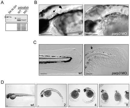
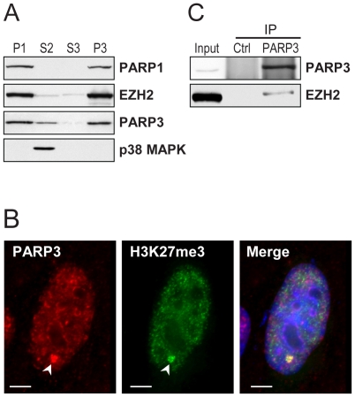
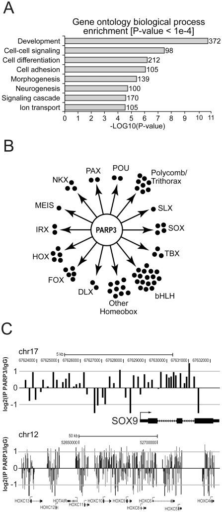
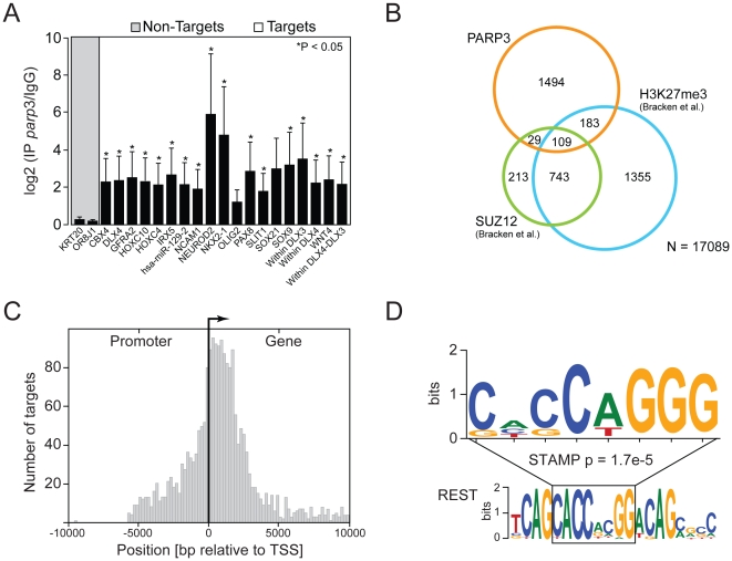
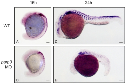
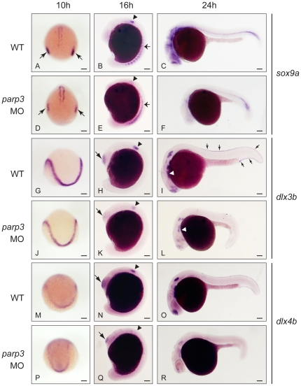
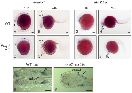
Principal findings: We have used several in vitro and in vivo approaches to examine the possible functions of PARP3 as a transcriptional regulator, a function suggested from its previously reported association with several Polycomb group (PcG) proteins. We demonstrate that PARP3 gene occupancy in the human neuroblastoma cell line SK-N-SH occurs preferentially with developmental genes regulating cell fate specification, tissue patterning, craniofacial development and neurogenesis. Addressing the significance of this association during zebrafish development, we show that morpholino oligonucleotide-directed inhibition of parp3 expression in zebrafish impairs the expression of the neural crest cell specifier sox9a and of dlx3b/dlx4b, the formation of cranial sensory placodes, inner ears and pectoral fins. It delays pigmentation and severely impedes the development of the median fin fold and tail bud.

Conclusion: Our findings demonstrate that Parp3 is crucial in the early stages of zebrafish development, possibly by exerting its transcriptional regulatory functions as early as during the specification of the neural plate border.


Conflict of interest statement

Competing Interests: The authors have declared that no competing interests exist.

Figures

Figure 1
Figure 1. Developmental perturbations in zebrafish embryos with impaired parp3 expression.
A. Immunoblot analysis (upper panel) of zebrafish Parp3 in wild type (WT) and parp3 morphants (MO) using an antibody raised against human PARP3. A whole cell extract of human SK-N-SH cells is shown as a control. The protein bands corresponding to PARP3 are indicated by “>”. The faster migrating band corresponds to a non-related protein that cross-reacts with the antibody. The Western blot membrane was stained with Ponceau S as a protein loading control (lower panel). B. Enlarged lateral views of the head regions of wild type and parp3 morphants injected with 4 ng MO1. The inner ears (small arrow) and the pectoral fins (large arrow) in the wt embryos are not formed in the parp3 morphants. C. Enlarged lateral views of the tail of wild type and parp3 morphants injected with 4 ng MO1. The median fin fold (arrow) is less developed in the morphants and has a more granular aspect. Effects are more pronounced on the dorsal side (arrow). D. Zebrafish embryos 48hrs after injection of increasing amounts of the parp3-specific morpholino oligonucleotide MO1 at the one-cell stage (ng amounts indicated in the lower right corner). The short length of morphant embryos, their curved tail and their reduced pigmentation is increasingly severe with increasing amounts of injected MO1. Lateral views with anterior to the right and dorsal to the top. Scale bars represent 10 µm.
Figure 2
Figure 2. PARP3 is associated with chromatin and Polycomb proteins.
A. The human neuroblastoma cell line SK-N-SH was fractionated into nuclear (P1) and cytoplasmic (S2) fractions. The nuclear P1 fraction was further separated into nuclear soluble (S3) and chromatin (P3) fractions. Proteins detected by immunoblotting are indicated. PARP1 and p38 MAPK were used as chromatin and cytoplasmic markers, respectively. Samples of each fraction correspond to an equal cell number. B. Immunofluorescence detection of PARP3 in SK-N-SH cells. PARP3 is predominantly nuclear and co-localizes with trimethylated histone H3K27 (H3K27me3) (arrowhead). Scale bars represent 3 µm. C. Immunoblot analysis of proteins immunoprecipitated (IP) with nuclear PARP3. EZH2 co-precipitates with PARP3. Input corresponds to 10% of the cellular extract used in IP. Rabbit IgG were used in the control (Ctrl) IP.
Figure 3
Figure 3. PARP3 targets developmental genes.
A. Enrichment of PARP3 targets according to gene ontology annotations. B. Families of development-related transcription factors targeted by PARP3, as identified by ChIP-chip analysis. C. PARP3 ChIP-chip significant signals at SOX9 and HOXC loci. Rabbit IgG were used for the control ChIP. Transcription start sites are indicated by an arrow.
Figure 4
Figure 4. Analysis of PARP3-bound targets.
A. Validation of PARP3 developmental targets by ChIP-qPCR. PARP3 ChIP were analyzed by standard qPCR using primers specific for regions targeted by PARP3 according to ChIP-chip results. Error bars represent the standard deviation from three independent experiments and asterisks indicate a significant enrichment relative to control (p<0.05). Two control regions (KRT20 and OR8J1), which are not bound by PARP3 (Non-targets), were used to determine basal signal. Several probes were used for DLX genes. The “DLX4” probe targets the promoter region, “within DLX” probes target sequences within the DLX genes and “within DLX3-DLX4” probe targets the DLX3-DLX4 intergenic region. B. Overlaps between PARP3 and SUZ12 targets or sequences enriched in H3K27me3. SUZ12 and H3K27me3 targets were those determined in human embryonic fibroblasts by . C. Distribution of PARP3 target sequences relative to transcriptional start sites (TSS). D. PARP3 target sequences are enriched for the sequence CACCAGGG (upper sequence). This sequence matches part of the RE1-silencing transcription factor (REST) binding sequence (lower sequence).
Figure 5
Figure 5. Expression of the neural crest cell marker crestin is impaired in parp3 morphants.
Zebrafish embryos were untreated (WT) or injected with 4 ng parp3 MO1 and crestin expression was monitored by in situ hybridization. A. In 16 hpf WT embryos, crestin is expressed in premigratory neural crest cells and in neural crest cells migrating in the anterior trunk segments. B. In 16 hpf parp3 morphants, crestin expression appears to be generally reduced. C. By 24 hpf, crestin-positive cells are distributed along the anterior-posterior axis, in neural crest migratory pathways of WT embryos. D. In 24 hpf parp3 morphants, crestin expression is no longer detectable in the head and is markedly reduced in the trunk. Lateral views of embryos are shown with anterior to the left and dorsal to the top. Scale bars represent 10 µm.
Figure 6
Figure 6. Impaired expression of sox9a, dlx3b and dlx4b in parp3 morphants.
Zebrafish embryos were untreated (WT) or were injected with 4 ng parp3 MO1. Gene expression was detected by in situ hybridization. A–F. The expression of sox9a is drastically reduced in the otic placodes (small arrows) at 10 hpf and in the otic vesicles (arrowheads) at 16 hpf. Expression of sox9a in somite cells (small arrows in B and E) appears diffuse in parp3 morphants. Expression of sox9a is almost completely abolished in the head region at 24 hpf (C, F). Expressions of dlx3b (G–L) and dlx4b (M–R) are minimally affected by parp3 MO in ectodermal cells at 10 hpf (G, J, M, P) but are significantly reduced in the otic vesicles (arrowheads), olfactory placodes (large arrows) and branchial arches (white arrows) of parp3 morphants at 16 hpf (H, K, N, Q) and 24 hpf (I, L, O, R). The expression of dlx3b and dlx4b is abolished in the median fin fold of 24 hpf parp3 morphant embryos (small arrows in I). Dorsal views of embryos with anterior to the bottom in A, D, G, J, M, P and lateral views with anterior to the left, dorsal to the top, in B, C, E, F, H, I, K, L, N, O, Q and R. Scale bars represent 10 µm.
Figure 7
Figure 7. Expression of neurod and nkx2.1 in parp3 morphants.
Expression of: A–F, neurod and G–J, nkx2.1a were determined in uninjected embryos or in embryos that received 4 ng of parp3 MO1, by in situ hybridization. A–D and G–J are lateral views with anterior to the left, dorsal to the top. E–F are dorsal views of flat-mounted embryos with anterior to the left. ad/av/f: anterodorsal/anteroventral lateral line/facial placodes/ganglia; p: posterior lateral line placode; e: eye; o: octavel/statosacoustic ganglia precursors; t: telencephalon. Scale bars represent 10 µm.

References

    1. Hassa PO, Hottiger MO. The diverse biological roles of mammalian PARPS, a small but powerful family of poly-ADP-ribose polymerases. Front Biosci. 2008;13:3046–3082. - PubMed
    1. Yelamos J, Schreiber V, Dantzer F. Toward specific functions of poly(ADP-ribose) polymerase-2. Trends Mol Med. 2008;14:169–178. - PubMed
    1. Johansson M. A human poly(ADP-ribose) polymerase gene family (ADPRTL): cDNA cloning of two novel poly(ADP-ribose) polymerase homologues. Genomics. 1999;57:442–445. - PubMed
    1. Augustin A, Spenlehauer C, Dumond H, Menissier-De Murcia J, Piel M, et al. PARP-3 localizes preferentially to the daughter centriole and interferes with the G1/S cell cycle progression. J Cell Sci. 2003;116:1551–1562. - PubMed
    1. Rouleau M, McDonald D, Gagne P, Ouellet ME, Droit A, et al. PARP-3 associates with polycomb group bodies and with components of the DNA damage repair machinery. J Cell Biochem. 2007;100:385–401. - PubMed

Publication types

MeSH terms