. 2011 Jan 14;6(1):e14525.
doi: 10.1371/journal.pone.0014525.

Regulation of endothelial cell adhesion molecule expression by mast cells, macrophages, and neutrophils

Affiliations

Regulation of endothelial cell adhesion molecule expression by mast cells, macrophages, and neutrophils

Jie Zhang et al. PLoS One. .

Abstract

Background: Leukocyte adhesion to the vascular endothelium and subsequent transendothelial migration play essential roles in the pathogenesis of cardiovascular diseases such as atherosclerosis. The leukocyte adhesion is mediated by localized activation of the endothelium through the action of inflammatory cytokines. The exact proinflammatory factors, however, that activate the endothelium and their cellular sources remain incompletely defined.

Methods and results: Using bone marrow-derived mast cells from wild-type, Tnf(-/-), Ifng(-/-), Il6(-/-) mice, we demonstrated that all three of these pro-inflammatory cytokines from mast cells induced the expression of vascular cell adhesion molecule-1 (VCAM-1), intercellular adhesion molecule-1 (ICAM-1), P-selectin, and E-selectin in murine heart endothelial cells (MHEC) at both mRNA and protein levels. Compared with TNF-α and IL6, IFN-γ appeared weaker in the induction of the mRNA levels, but at protein levels, both IL6 and IFN-γ were weaker inducers than TNF-α. Under physiological shear flow conditions, mast cell-derived TNF-α and IL6 were more potent than IFN-γ in activating MHEC and in promoting neutrophil adhesion. Similar observations were made when neutrophils or macrophages were used. Neutrophils and macrophages produced the same sets of pro-inflammatory cytokines as did mast cells to induce MHEC adhesion molecule expression, with the exception that macrophage-derived IFN-γ showed negligible effect in inducing VCAM-1 expression in MHEC.

Conclusion: Mast cells, neutrophils, and macrophages release pro-inflammatory cytokines such as TNF-α, IFN-γ, and IL6 that induce expression of adhesion molecules in endothelium and recruit of leukocytes, which is essential to the pathogenesis of vascular inflammatory diseases.


Conflict of interest statement

Competing Interests: The authors have declared that no competing interests exist.

Figures

Figure 1
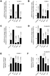
Figure 1. Degranulated mast cells induced MHEC adhesion molecule expression.
RT-PCR determined VCAM-1 (A), ICAM-1 (B), P-selectin (C), E-selectin (D), ICAM-1 (E), and P-selectin (F) mRNA levels relative to β-actin in MHEC after stimulation with conditioned media of mast cells from WT and different cytokine-deficient mice, or WT mast cell conditioned media pre-treated with anti-mouse TNF-α antibody or isotype control IgG (R&D Systems). Fold change was calculated relative to unstimulated MHEC. Data were presented as mean ± SE of three independent experiments. P<0.05 was considered statistically significant, Student's t test. *Compared with WT BMMC-treated MHEC.
Figure 2
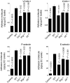
Figure 2. Adhesion molecule protein levels from MHEC treated with different degranulated mast cell supernatants.
Commercial ELISA kits were used to determine VCAM-1, ICAM-1, P-selectin, and E-selectin in MHEC lysate. Degranulated mast cell supernatants from different mice, as indicated, were used to treat mouse MHEC for 24 hours. Data are presented as mean ± SE of three independent experiments. P<0.05 was considered statistically significant, Student's t test. *Compared with WT BMMC-treated MHEC.
Figure 3
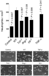
Figure 3. Neutrophil adhesion assay on activated MHEC monolayers under physiological shear stress.
MHEC monolayer was activated with different degranulated mast cell supernatants for 16 hours and then examined for adhesion of bone marrow neutrophils from WT mice under shear flow conditions of 0.5 dynes/cm2. Neutrophil adhesion on MHEC monolayers was quantified as reported previously. Culture media alone was used as a negative control and recombinant TNF-α (25 ng/mL) was used as a positive control. Data were the mean ± SE from eight different fields. P<0.05 was considered statistically significant, Student's t test. *Compared with WT BMMC-treated MHEC. Representative photographs captured from videos are shown in the bottom panels. Arrows indicate examples of adhered cells bound to MHEC. Cells that are not adhered to MHEC are flowing by, therefore they are out of focus and not as bright as adhered cells: Examples are indicated with #.
Figure 4
Figure 4. Adhesion molecule expression in MHEC after macrophage stimulation.
MHEC were treated with macrophage cell lysates from different mice, as indicated, for four hours. VCAM-1 (A), ICAM-1 (B), P-selectin (C), and E-selectin (D) mRNA levels were quantified by RT-PCR and normalized to β-actin. Fold changes were calculated relative to unstimulated EC. ELISA determined VCAM-1 (E) and E-selectin (F) protein levels in lysates of MHEC that were stimulated with macrophage lysates from different mice for 24 hours. Data were presented as mean ± SE of three to six independent experiments. P<0.05 was considered statistically significant, Student's t test. *Compared with MHEC treated with WT macrophage lysate.
Figure 5
Figure 5. Adhesion molecule expression in MHEC after neutrophil stimulation.
Neutrophil lysates were prepared from bone marrow from different mice, as indicated, and used to induce MHEC for four hours. VCAM-1 (A), ICAM-1 (B), P-selectin (C), and E-selectin (D) mRNA levels were quantified by RT-PCR and normalized to β-actin. Fold changes were determined relative to unstimulated MHEC. ELISA determined VCAM-1 (E) and E-selectin (F) protein levels in lysates of MHEC that were stimulated with neutrophil lysates from different mice, as indicated, for 24 hours. Data were presented as mean ± SE of three to six independent experiments. P<0.05 was considered statistically significant, Student's t test. *Compared with MHEC treated with WT neutrophil lysate.

References

    1. Vestweber D, Blanks JE. Mechanisms that regulate the function of the selectins and their ligands. Physiol Rev. 1999;79:181–213. - PubMed
    1. Varki A. Selectin ligands. Proc Natl Acad Sci USA. 1994;91:7390–7397. - PMC - PubMed
    1. Collins RG, Velji R, Guevara NV, Hicks MJ, Chan L, et al. P-Selectin or intercellular adhesion molecule (ICAM)-1 deficiency substantially protects against atherosclerosis in apolipoprotein E-deficient mice. J Exp Med. 2000;191:189–194. - PMC - PubMed
    1. Takahashi K, Takeya M, Sakashita N. Multifunctional roles of macrophages in the development and progression of atherosclerosis in humans and experimental animals. Med Electron Microsc. 2002;35:179–203. - PubMed
    1. Johnson-Tidey RR, McGregor JL, Taylor PR, Poston RN. Increase in the adhesion molecule P-selectin in endothelium overlying atherosclerotic plaques. Coexpression with intercellular adhesion molecule-1. Am J Pathol. 1994;144:952–961. - PMC - PubMed

Publication types