. 2011 Feb 1;71(3):925-36.
doi: 10.1158/0008-5472.CAN-10-1032. Epub 2011 Jan 25.

Phosphorylation regulates c-Myc's oncogenic activity in the mammary gland

Affiliations

Phosphorylation regulates c-Myc's oncogenic activity in the mammary gland

Xiaoyan Wang et al. Cancer Res. .

Abstract

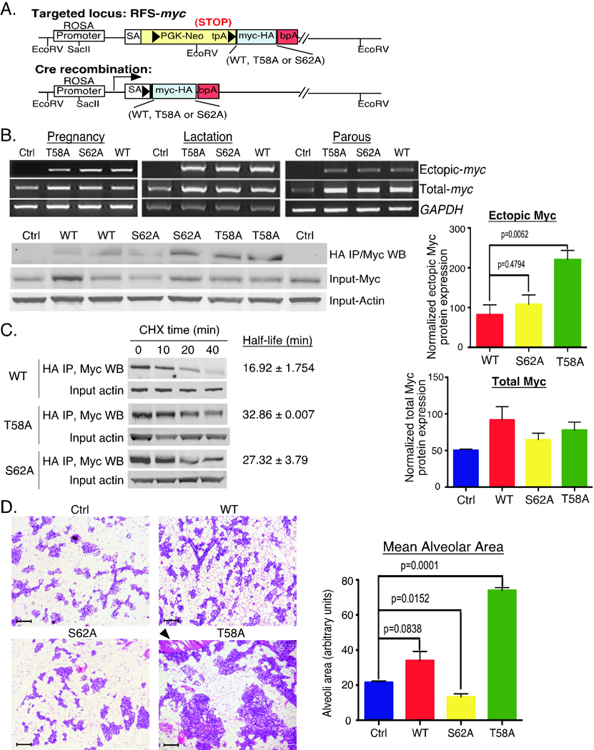
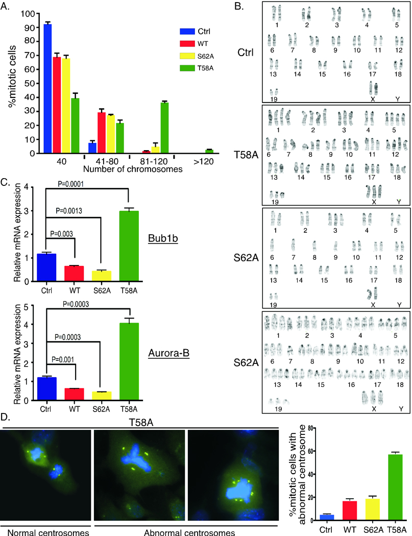
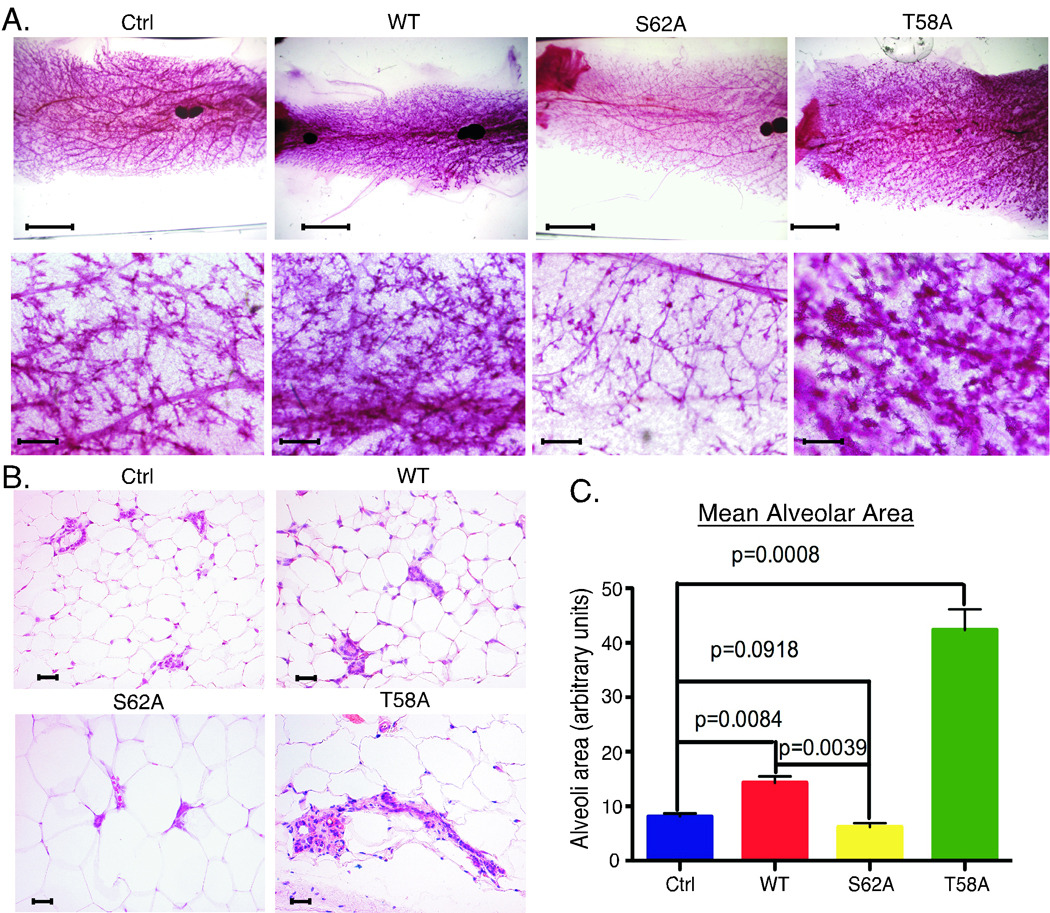
Expression of the c-Myc oncoprotein is affected by conserved threonine 58 (T58) and serine 62 (S62) phosphorylation sites that help to regulate c-Myc protein stability, and altered ratios of T58 and S62 phosphorylation have been observed in human cancer. Here, we report the development of 3 unique c-myc knock-in mice that conditionally express either c-Myc(WT) or the c-Myc(T58A) or c-Myc(S62A) phosphorylation mutant from the constitutively active ROSA26 locus in response to Cre recombinase to study the role of these phosphorylation sites in vivo. Using a mammary-specific Cre model, we found that expression of c-Myc(WT) resulted in increased mammary gland density, but normal morphology and no tumors at the level expressed from the ROSA promoter. In contrast, c-Myc(T58A) expression yielded enhanced mammary gland density, hyperplastic foci, cellular dysplasia, and mammary carcinoma, associated with increased genomic instability and suppressed apoptosis relative to c-Myc(WT). Alternatively, c-Myc(S62A) expression reduced mammary gland density relative to control glands, and this was associated with increased genomic instability and normal apoptotic function. Our results indicate that specific activities of c-Myc are differentially affected by T58 and S62 phosphorylation. This model provides a robust platform to interrogate the role that these phosphorylation sites play in c-Myc function during development and tumorigenesis.


Conflict of interest statement

Conflict of Interests

The authors declare that they have no conflict of interest.

Figures

Figure 1
Figure 1. Generation and characterization of RFS-mycWT/WAP-Cre, RFS-mycT58A/WAP-Cre and RFS-mycS62A/WAP-Cre mice
A. Knock-in strategy for conditional expression of c-mycWT-HA, c-mycT58A-HA or c-mycS62A-HA. Arrowheads represent loxP sites. tpA is a transcription stop sequence. Inserted c-myc cDNAs have a C-terminal HA tag. Cre recombination activates expression of the inserted c-myc cDNA driven from the ROSA promoter. RFS=ROSA Floxed Stop (see Suppl. Fig. 1 for more detail). B. Expression of ROSA-driven c-Myc. Upper panels: RNA was isolated from mammary glands from the indicated control (ctrl: no Wap-Cre) or RFS-myc/WAP-Cre strains (WT, T58A, S62A) at the indicated stages: pregnancy 18 days, lactation 18 days, parous 2 months. HA-tagged ectopic and total c-myc mRNA is shown by RT-PCR. Lower panels: lysates from mammary glands from 2 mice per indicated strain harvested at pregnancy day 17 were immunoprecipitated with anti-HA followed by western blotting with anti-c-Myc. Input total c-Myc and Actin are shown. Data is representative of 4 mice per genotype. Graphs: quantification of the Actin-normalized expression of HA-tagged ectopic and total c-Myc from 3 mice per genotype ± SD. P-values are given on graphs with significant differences. C. Analysis of ectopic c-Myc half-life in primary MECs from the indicated strains. Mean half-life ± SD was calculated from three independent experiments. D. Expanded lobule-alveolar areas with c-MycT58A expression. The 4th glands from the indicated mice on day 17 of the 3rd consecutive pregnancy were analyzed by H&E section staining. Arrowhead indicates enlarged duct. Scale bars are 50 µM. Data is representative of 4 mice per genotype. Mean alveolar area was analyzed by counting 10 areas each section and 3 mice per genotype by ImageJ and graphed ± SD.
Figure 2
Figure 2. Inhibition of mammary gland involution and apoptosis with c-MycT58A expression
A. The 4th gland from the indicated mice was harvested after 12 days of lactation, 3 days post weaning after the 3rd pregnancy, and analyzed by H&E section staining. Scale bars are 50µM. Data is representative of 4–6 mice per genotype. B. c-MycT58A expression inhibits apoptosis during involution. Mammary gland sections as in A were analyzed by TUNEL assay. Scale bars are 50µM. C. TUNEL assays as in B were quantified to determine the percent apoptotic epithelial cells. Graph represents 4 mice per strain ± SD. D. Reduced expression of pro-apoptotic Bim in mammary glands expressing c-MycT58A. Protein lysates from the indicated mice (2 per genotype) 3 days post weaning were western blotted with the indicated antibodies and quantified. Data is representative of 4 mice per genotype. Average Actin-normalized BimEL and BimML expression relative to control is graphed ± SD.
Figure 3
Figure 3. Increased genomic instability with expression of c-Myc phosphorylation mutants
A. Mitotic spreads from primary MECs from the indicated strains were analyzed for chromosome numbers. Average percent mitotic cells with diploid (40 chromosomes) and hyperdiploid karyotypes from 3 mice per strain ± SD is graphed. B. Expression of c-MycT58A and c-MycS62A induces aneuploidy. Representative G-banding of mitotic cells from control mice or mice expressing c-MycT58A or c-MycS62A are shown. Cells were prepared as in A. Twenty mitotic cells were analyzed per strain. C. Substantial and opposing changes in expression of spindle checkpoint genes with c-MycT58A and c-MycS62A expression. RNA was isolated from 2-month parous mammary glands from the indicated strains. Quantitative RT-PCR analysis for the expression of Bub1b and Aurora-B genes relative to control mice are graphed ± SD. Data represents 4–5 mice per strain. D. Substantial centrosome amplification with expression of c-MycT58A. Centrosomes were visualized by anti-pericentrin immunofluorescence staining (green) and DNA by DAPI (blue). Cells were prepared as in A. Representative MycT58A cells are shown. Centrosome amplification (more than 2 per mitotic cell) was quantified from three mice per indicated strains and graphed ± SD.
Figure 4
Figure 4. Characterization of mammary glands expressing c-MycWT, c-MycT58A and c-MycS62A at two months parous
A. The 4th glands from the indicated strains of mice two months parous after the 3rd pregnancy were analyzed by whole mount. Solid, darkly staining areas are lymph nodes. Scale bars are 5 mM (top row) and 200 µM (bottom row). Images shown are representative of 3–4 mice per genotype. B. H&E sections from mice as in A. Scale bars are 50 µM. Images are representative of 4 mice per genotype. C. Expanded lobule-alveolar areas with c-MycT58A expression. Mean alveolar area from mice as in A was analyzed by counting 10 areas each H&E section. 3 mice per genotype ± SD is graphed.
Figure 5
Figure 5. Preneoplastic changes with expression of c-MycT58A at five to eight months parous
A. Hyperplastic foci in RFS-mycT58A/WAP-Cre mammary glands 5–8 months parous after the 3rd pregnancy. Whole mount, H&E section, and IHC for BrdU labelling are shown. Arrow in whole mount indicates hyperplastic foci. In H&E, dark blue/purple represents calcifications in acinar lumens. B. Areas of cellular dysplasia in MycT58A mammary glands. H&E sections from MycT58A mice as in A. Arrows (from left to right) indicate atypical nuclei with multiple nucleoli, mitotic figure, and mast cell. C. Alveolar disorganization and reduced adhesion in MycT58A mammary glands. Immunofluorescence co-staining for luminal marker K8/18 (green) and myoepithelial marker K14 (red) in mice as in A. Arrow indicates disorganized alveolar region. E-cadherin staining (red), Dapi stained nuclei (blue) in MycT58A (middle panel) and MycWT (right panel) mammary glands. D. Ductal hyperplasia with c-MycT58A expression. H&E staining and immunofluorescence K8/18 (green)/K14 (red) in mice as in A. Dilated ducts with proliferative epithelium (tufting) are shown. All whole mount and H&E images are from different MycT58A mice and are representative of 8 RFS-mycT58A/WAP-Cre mice 5–8 months parous. Immunofluorescence images are representative of 3 mice per genotype. All scale bars are 50µM.
Figure 6
Figure 6. Expression of c-MycT58A induces mammary adenocarcinoma with an average latency of 9 months
A. Examples of mammary tumors in MycT58A expressing glands. Gross photograph of 5th gland tumor in 10-month old RFS-mycT58A/Wap-Cre mouse (upper left), H&E section showing the cribriform architecture of same (upper right), adenosquamous tumor from 7-month RFS-mycT58A/Wap-Cre mouse (lower left), adenocarcinoma with solid architecture in different 10-month RFS-mycT58A/Wap-Cre mouse (lower right). B. High proliferation in MycT58A-driven tumor. Ki-67 staining of tumor shown in A, lower right. C. Invasive features of tumor from MycT58A gland. Immunohistochemistry for smooth muscle actin (SMA) in normal duct (upper panel) close to the periphery of tumor shown in A, upper right, and loss of SMA expression in the tumor (lower panel). D. Metastasis to liver and lung of MycT58A-induced mammary tumor shown in A, lower right. All scale bars are 50 µM.

References

    1. Meyer N, Penn LZ. Reflecting on 25 years with MYC. Nat Rev Cancer. 2008;8:976–990. - PubMed
    1. Nesbit CE, Tersak JM, Prochownik EV. MYC oncogenes and human neoplastic disease. Oncogene. 1999;18:3004–3016. - PubMed
    1. D'Cruz CM, Gunther EJ, Boxer RB, et al. c-MYC induces mammary by means of a preferred pathway involving spontaneous Kras2 mutations. Nat Med. 2001;7:235–239. - PubMed
    1. Pelengaris S, Khan M, Evan G. c-MYC: more than just a matter of life and death. Nat Rev Cancer. 2002;2:764–776. - PubMed
    1. Khan M, Evan G, Pelengaris S. Suppression of Myc-induced apoptosis in beta cells exposes multiple oncogenic properties of Myc and triggers carcinogenic progression. Nature Reviews Cancer. 2002;2:764–776. - PubMed

Publication types

MeSH terms

Substances