

DeltaNp63alpha confers tumor cell resistance to cisplatin through the AKT1 transcriptional regulation
- PMID: 21266360
- PMCID: PMC3076926
- DOI: 10.1158/0008-5472.CAN-10-1481
DeltaNp63alpha confers tumor cell resistance to cisplatin through the AKT1 transcriptional regulation
Abstract
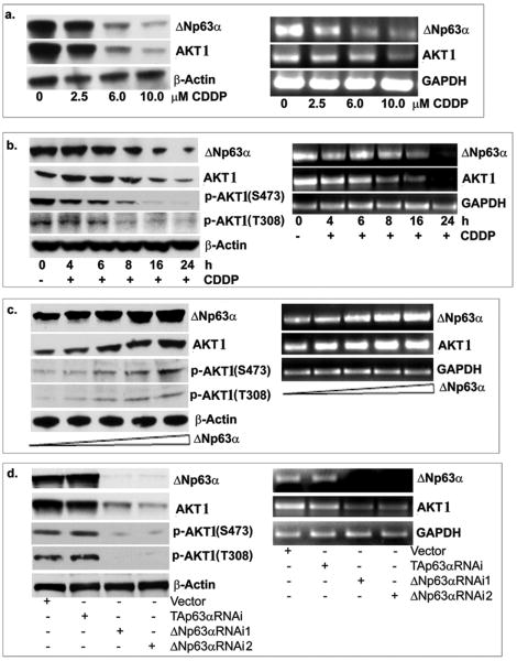
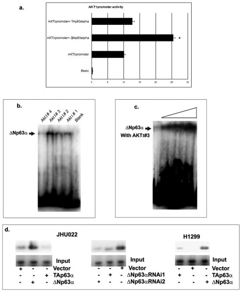
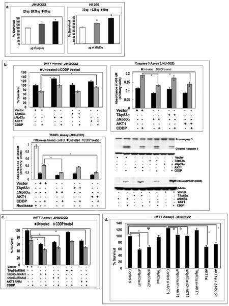
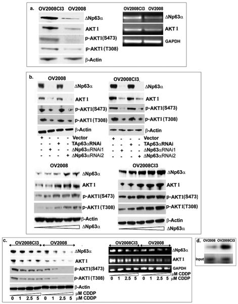
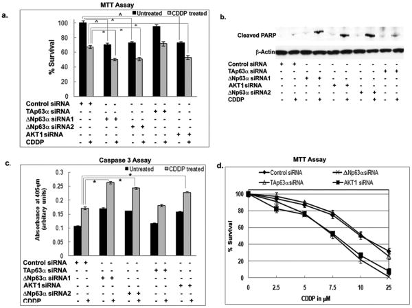
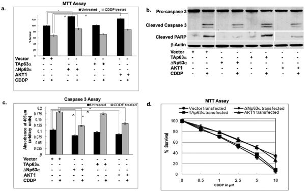
Strategies to address resistance to platin drugs are greatly needed in human epithelial cancers (e.g., ovarian, head/neck, and lung) where platins are used widely and resistance occurs commonly. We found that upon ΔNp63α overexpression, AKT1 and phospho-AKT1 levels are upregulated in cancer cells. Investigations using gel-shift, chromatin immunoprecipitation and functional reporter assays implicated ΔNp63α in positive regulation of AKT1 transcription. Importantly, we found that ΔNp63α, AKT1, and phospho-AKT levels are greater in 2008CI3 CDDP-resistant ovarian cancer cells than in 2008 CDDP-sensitive cells. siRNA-mediated knockdown of ΔNp63α expression dramatically decreased AKT1 expression, whereas knockdown of either ΔNp63α or AKT1 decreased cell proliferation and increased death of ovarian and head/neck cancer cells. Conversely, enforced expression of ΔNp63α increased cancer cell proliferation and reduced apoptosis. Together, our findings define a novel ΔNp63α-dependent regulatory mechanism for AKT1 expression and its role in chemotherapeutic resistance of ovarian and head/neck cancer cells.
Figures
Similar articles
-
Phospho-DeltaNp63alpha/NF-Y protein complex transcriptionally regulates DDIT3 expression in squamous cell carcinoma cells upon cisplatin exposure.Cell Cycle. 2010 Jan 15;9(2):328-38. doi: 10.4161/cc.9.2.10432. Epub 2010 Jan 26. Cell Cycle. 2010. PMID: 20023394
-
Phospho-ΔNp63α/SREBF1 protein interactions: bridging cell metabolism and cisplatin chemoresistance.Cell Cycle. 2012 Oct 15;11(20):3810-27. doi: 10.4161/cc.22022. Epub 2012 Sep 5. Cell Cycle. 2012. PMID: 22951905 Free PMC article.
-
U-box-type ubiquitin E4 ligase, UFD2a attenuates cisplatin mediated degradation of DeltaNp63alpha.Cell Cycle. 2008 May 1;7(9):1231-7. doi: 10.4161/cc.7.9.5795. Epub 2008 Feb 19. Cell Cycle. 2008. PMID: 18418053 Free PMC article.
-
Phospho-ΔNp63α/miR-885-3p axis in tumor cell life and cell death upon cisplatin exposure.Cell Cycle. 2011 Nov 15;10(22):3938-47. doi: 10.4161/cc.10.22.18107. Epub 2011 Nov 15. Cell Cycle. 2011. PMID: 22071691 Free PMC article.
-
Molecular Mechanisms of p63-Mediated Squamous Cancer Pathogenesis.Int J Mol Sci. 2019 Jul 23;20(14):3590. doi: 10.3390/ijms20143590. Int J Mol Sci. 2019. PMID: 31340447 Free PMC article. Review.
Cited by
-
1α, 25-Dihydroxyvitamin D₃ and the vitamin D receptor regulates ΔNp63α levels and keratinocyte proliferation.Cell Death Dis. 2015 Jun 11;6(6):e1781. doi: 10.1038/cddis.2015.148. Cell Death Dis. 2015. PMID: 26068789 Free PMC article.
-
ΔNp63α exerts antitumor functions in cervical squamous cell carcinoma.Oncogene. 2020 Jan;39(4):905-921. doi: 10.1038/s41388-019-1033-x. Epub 2019 Oct 1. Oncogene. 2020. PMID: 31576015
-
Inhibition of USP28 overcomes Cisplatin-resistance of squamous tumors by suppression of the Fanconi anemia pathway.Cell Death Differ. 2022 Mar;29(3):568-584. doi: 10.1038/s41418-021-00875-z. Epub 2021 Oct 5. Cell Death Differ. 2022. PMID: 34611298 Free PMC article.
-
Analysis of apoptosis methods recently used in Cancer Research and Cell Death & Disease publications.Cell Death Dis. 2012 Feb 2;3(2):e263. doi: 10.1038/cddis.2012.2. Cell Death Dis. 2012. PMID: 22297295 Free PMC article. No abstract available.
-
Common Chromosomal Fragile Site Gene WWOX in Metabolic Disorders and Tumors.Int J Biol Sci. 2014 Jan 11;10(2):142-8. doi: 10.7150/ijbs.7727. eCollection 2014. Int J Biol Sci. 2014. PMID: 24520212 Free PMC article. Review.
References
-
- Kelland L. The resurgence of platinum-based cancer chemotherapy. Nat Rev Cancer. 2007;7:573–84. - PubMed
-
- Helmbach H, Kern MA, Rossmann E, et al. Drug resistance towards etoposide and cisplatin in human melanoma cells is associated with drug-dependent apoptosis deficiency. J Invest Dermatol. 2002;118:923–32. - PubMed
-
- Dan HC, Sun M, Kaneko S, et al. Akt phosphorylation and stabilization of X-linked inhibitor of apoptosis protein (XIAP) J Biol Chem. 2004;279:5405–12. - PubMed
-
- Fraser M, Leung BM, Yan X, et al. p53 is a determinant of X-linked inhibitor of apoptosis protein/Akt-mediated chemoresistance in human ovarian cancer cells. Cancer Res. 2003;63:7081–8. - PubMed
-
- Page C, Lin HJ, Jin Y, et al. Overexpression of Akt can modulate chemotherapy-induced apoptosis. Anticancer Res. 2000;20:407–16. - PubMed
Publication types
MeSH terms
Substances
Grants and funding
LinkOut - more resources
Full Text Sources
Miscellaneous
