. 2011 Feb;138(4):653-65.
doi: 10.1242/dev.056499.

Sox9+ ductal cells are multipotent progenitors throughout development but do not produce new endocrine cells in the normal or injured adult pancreas

Affiliations

Sox9+ ductal cells are multipotent progenitors throughout development but do not produce new endocrine cells in the normal or injured adult pancreas

Janel L Kopp et al. Development. 2011 Feb.

Abstract

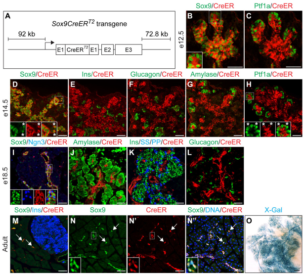
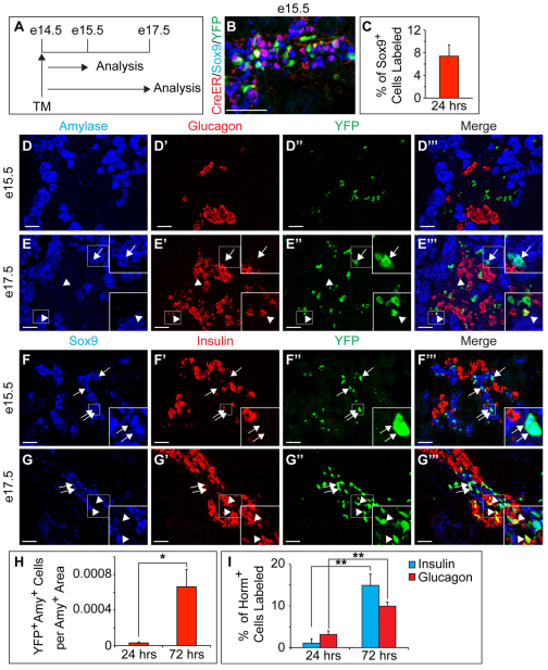
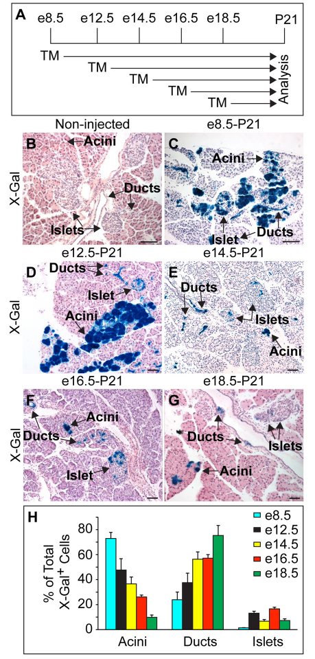
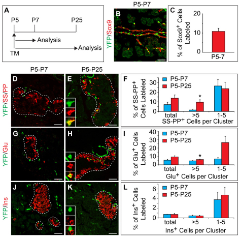
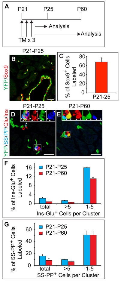
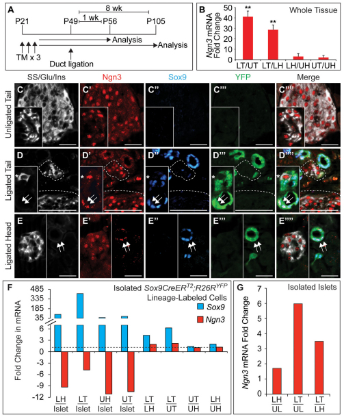
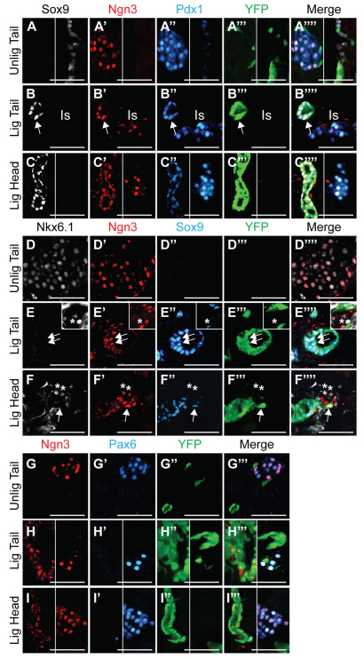
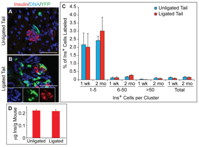
One major unresolved question in the field of pancreas biology is whether ductal cells have the ability to generate insulin-producing β-cells. Conclusive examination of this question has been limited by the lack of appropriate tools to efficiently and specifically label ductal cells in vivo. We generated Sox9CreER(T2) mice, which, during adulthood, allow for labeling of an average of 70% of pancreatic ductal cells, including terminal duct/centroacinar cells. Fate-mapping studies of the Sox9(+) domain revealed endocrine and acinar cell neogenesis from Sox9(+) cells throughout embryogenesis. Very small numbers of non-β endocrine cells continue to arise from Sox9(+) cells in early postnatal life, but no endocrine or acinar cell neogenesis from Sox9(+) cells occurs during adulthood. In the adult pancreas, pancreatic injury by partial duct ligation (PDL) has been suggested to induce β-cell regeneration from a transient Ngn3(+) endocrine progenitor cell population. Here, we identify ductal cells as a cell of origin for PDL-induced Ngn3(+) cells, but fail to observe β-cell neogenesis from duct-derived cells. Therefore, although PDL leads to activation of Ngn3 expression in ducts, PDL does not induce appropriate cues to allow for completion of the entire β-cell neogenesis program. In conclusion, although endocrine cells arise from the Sox9(+) ductal domain throughout embryogenesis and the early postnatal period, Sox9(+) ductal cells of the adult pancreas no longer give rise to endocrine cells under both normal conditions and in response to PDL.


Figures

Fig. 1.
Fig. 1.
Sox9CreERT2 specifically and efficiently labels Sox9+ cells in mouse pancreas. (A) Schematic of the Sox9CreERT2 transgene. (B-N′) Double and triple immunofluorescent labeling of CreER and various hormones and markers at e12.5, e14.5, e18.5 and P25 (adult). Immunofluorescence analysis for Cre recombinase (CreER) shows specific and uniform expression of CreER in the Sox9+ domain. Insets and asterisks in B, D and I show Sox9+ CreER+ cells (magnification of boxed areas). CreER expression is absent from hormone+ (E,F,K,L,M) and amylase+ cells (G,J). CreER expression recedes from Ptf1a+ tips between e12.5 (C) and e14.5 (H, asterisks in inset show CreER+ Ptf1a+ cells). Forty-eight hours after three tamoxifen (TM) injections at P21, CreER is detected in the nucleus (N-N′; insets show magnifications of boxed areas). Arrows in M-N′ point to Sox9/CreER coexpressing terminal duct/centroacinar cells. (O) Whole-mount β-galactosidase staining (X-Gal) of Sox9CreERT2;R26RlacZ mice injected with TM at P10 shows labeling of the entire ductal tree. Scale bars: 50 μm in B-N′; 500 μm in O. Ins, insulin; PP, pancreatic polypeptide; SS, somatostatin.
Fig. 2.
Fig. 2.
A Sox9+ Ptf1a+ Nkx6.1+ Hnf1b domain marks the distal ends of the embryonic cords of mouse pancreas. (A-A′) Immunofluorescence staining reveals that the Sox9+ and Hnf1b+ domains largely coincide at e14.5. However, some Sox9+ cells are Hnf1b (arrows). (B-B′′) At e14.5, cells coexpress Sox9, Ptf1a and Nkx6.1 (arrows). (C-C′′) Ptf1a+ Nkx6.1+ cells in this location do not express Hnf1b (arrows). (D) Schematic of different domains in the e14.5 pancreas. Scale bars: 50 μm.
Fig. 3.
Fig. 3.
Sox9+ embryonic cords give rise to endocrine and acinar cells in mouse pancreas. (A) Schematic of the lineage tracing experiment in Sox9CreERT2;R26RYFP mice. Tamoxifen (TM) was administered at e14.5 and pancreatic tissue was analyzed at e15.5 and e17.5. (B) Nuclear Cre recombinase (CreER) coincides with YFP in the Sox9+ domain. (C) Quantification of YFP+ Sox9+ cells relative to the total number of Sox9+ cells (n=3). (D-G′′) Immunofluorescence staining for amylase, glucagon and YFP (D-D′′,E-E′′) or Sox9, insulin and YFP (F-F′′,G-G′′). Arrows in E-E′′ indicate YFP+ amylase+ cells, arrowheads in E-E′′ YFP+ glucagon+ cells and arrowheads in G-G′′ YFP+ insulin+ cells. Arrows in F-F′′ and G-G′′ point to insulin YFP+ Sox9+ cells. Insets are magnified views of the boxed areas. (H,I) Quantification of YFP+ amylase+ cells relative to total amylase+ area (H) or insulin+ or glucagon+ cells relative to the number of total insulin+ or glucagon+ cells (I), respectively (n=3). Values are shown as mean ± s.e.m. *P<0.05; **P<0.01; Amy, amylase; horm, hormone. Scale bars: 50 μm.
Fig. 4.
Fig. 4.
Endocrine and acinar cells arise from the Sox9+ domain throughout development. (A) Schematic of the lineage tracing experiment in Sox9CreERT2;R26RlacZ mice. Tamoxifen (TM) was administered at various times during embryonic development and pancreatic tissue was analyzed at P21. (B-G) Without TM, no β-galactosidase staining (X-Gal) is detected (B). TM injection results in β-galactosidase staining (blue) of acini, ducts and islets at P21 (C-G). (H) β-galactosidase+ cells were quantified as a percentage of all β-galactosidase + cells (labeled as X-Gal+; n=3) in acini, ducts and islets. Values are shown as mean ± s.e.m. Scale bars: 50 μm.
Fig. 5.
Fig. 5.
Small numbers of non-β endocrine cells arise from Sox9+ ducts during the early postnatal period. (A) Schematic of the lineage tracing experiment in Sox9CreERT2;R26RYFP mice. Tamoxifen (TM) was administered at P5 and pancreatic tissue was analyzed at P7 and P25. (B) The YFP lineage label is detected primarily in Sox9+ cells. (C) Quantification of YFP+ Sox9+ cells relative to the total number of Sox9+ cells (n=3). (D,E,G,H) Immunofluorescence staining reveals YFP+ SS+ PP+ cells (insets in E) and YFP+ Glu+ cells (insets in H) located in islets (delineated by dashed line). (J,K) YFP does not colocalize with insulin (Ins) in islets (insets in K). (F,I,L) Quantification of YFP-labeled SS+ PP+ (F), Glu+ (I) or Ins+ (L) cells relative to the total number of SS+ PP+, Glu+ or Ins+ cells, respectively (n=3). Values are grouped according to endocrine cell cluster sizes. Values are shown as mean ± s.e.m. *P<0.05. Scale bars: 50 μm. Glu, glucagon; Ins, insulin; PP, pancreatic polypeptide; SS, somatostatin.
Fig. 6.
Fig. 6.
Endocrine cells do not arise from Sox9+ ducts during adulthood. (A) Schematic of the lineage tracing experiment in Sox9CreERT2;R26RYFP mice. Tamoxifen (TM) was administered daily for three days beginning at postnatal day P21 and pancreatic tissue was analyzed at P25 and P60. (B) The YFP lineage label is primarily detected in Sox9+ cells. (C) Quantification of YFP+ Sox9+ cells relative to the total number of Sox9+ cells (n=3). (D,E) Immunofluorescence staining reveals YFP+ SS+ PP+ cells and YFP+ Ins+ Glu+ cells closely associated with YFP+ ducts at P25 (insets in D) and P60 (insets in E). Asterisks indicate YFP+ hormone+ cells. (F,G) Quantification of YFP-labeled Ins+ Glu+ (F) or SS+ PP+ (G) cells relative to the total number of Ins+ Glu+ or SS+ PP+ cells, respectively (n=3). Values are grouped according to endocrine cell cluster sizes. Values are shown as mean ± s.e.m. Scale bars: 50 μm. Glu, glucagon; Ins, insulin; PP, pancreatic polypeptide; SS, somatostatin.
Fig. 7.
Fig. 7.
Ngn3 expression is induced in duct-derived cells and islets after partial duct ligation (PDL). (A) Schematic of the lineage tracing experiment in Sox9CreERT2;R26RYFP mice. Tamoxifen (TM) was administered daily for three days beginning at P21, duct ligation or sham-operation was performed at P49 and pancreatic tissue was analyzed one or eight weeks later. (B) Quantitative RT-PCR analysis of pancreatic heads and tails isolated from mice one week after sham-operation or PDL (n=5). (C-E′′) Immunofluorescence staining for somatostatin (SS), glucagon (Glu) and insulin (Ins) together with Ngn3, Sox9 and YFP reveals Ngn3 in endocrine cells in pancreatic tails of sham-operated mice (C-C′′) and tails (D-D′′, dashed lines delineate endocrine cell clusters) and heads (E-E′′) of mice seven days after PDL. Hormone+ Ngn3+ cells in ducts are YFP Sox9 (asterisks in D-D′′). After PDL, Ngn3 is expressed in YFP+ Sox9+ cells of the ligated tail (arrows in D-D′′) and head (arrows in E-E′′). (F,G) Quantitative RT-PCR analysis of YFP+ cells (F, n=4) or islets (G, n=6) isolated from mice one week after sham-operation or PDL. Values are shown as mean ± s.e.m. **P<0.01. LH, ligated head; LT, ligated tail; UH, unligated head; UT, unligated tail; UL, unligated whole pancreas. Scale bars: 50 μm.
Fig. 8.
Fig. 8.
Duct-derived Ngn3+ Sox9+ cells after partial duct ligation (PDL) express Pdx1 but not Nkx6.1 and Pax6. (A-I′′) Immunofluorescence staining of pancreata from PDL-treated or sham-operated Sox9CreERT2;R26RYFP mice reveals cells expressing Ngn3 together with Pdx1 (A-C′′), Nkx6.1 (D-F′′) or Pax6 (G-I′′) in pancreatic tails of sham-operated (A-A′′,D-D′′,G-G′′) and tails (B-B′′,E-E′′,H-H′′) and heads (C-C′′,F-F′′,I-I′′) of PDL-treated mice one week after surgery. A subset of YFP+ Sox9+ cells in tails of sham-operated mice express Pdx1 (A-A′′) but not Nkx6.1 (D-D′′) or Pax6 (G-G′′). Ngn3+ YFP Sox9 cells are also found in the pancreatic tails and heads of PDL-treated mice (asterisks in E-F′′ mark Ngn3+ Nkx6.1+ YFP Sox9 cells). Ngn3+ YFP+ Sox9+ cells after PDL express Pdx1 (arrows in B-B′′) but not Nkx6.1 (arrows in E-F′′) or Pax6 (H-I′′). Unlig Tail, unligated tail; Lig Tail, ligated tail; Lig Head, ligated head; Is, islet. Scale bars: 50 μm.
Fig. 9.
Fig. 9.
Sox9+ cells do not give rise to insulin+ cells after partial duct ligation (PDL). (A,B) Immunofluorescence analysis of pancreatic tails from PDL-treated or sham-operated Sox9CreERT2;R26RYFP mice for insulin and YFP. (C) Quantification of YFP+ insulin+ cells relative to the total number of insulin+ cells shows no increase in the percentage of YFP+ β-cells one week (1 wk; n=4) or two months (2 mo; n=3) after PDL. Values are shown for all insulin+ cells (total) and for insulin+ cell clusters of different sizes. (D) Whole pancreas insulin content relative to body weight (n=10). Values are shown as mean ± s.e.m. Ins, insulin. Scale bars: 50 μm.

References

    1. Akiyama H., Kim J. E., Nakashima K., Balmes G., Iwai N., Deng J. M., Zhang Z., Martin J. F., Behringer R. R., Nakamura T., et al. (2005). Osteo-chondroprogenitor cells are derived from Sox9 expressing precursors. Proc. Natl. Acad. Sci. USA 102, 14665-14670 - PMC - PubMed
    1. Blaine S. A., Ray K. C., Anunobi R., Gannon M. A., Washington M. K., Means A. L. (2010). Adult pancreatic acinar cells give rise to ducts but not endocrine cells in response to growth factor signaling. Development 137, 2289-2296 - PMC - PubMed
    1. Cheung M., Briscoe J. (2003). Neural crest development is regulated by the transcription factor Sox9. Development 130, 5681-5693 - PubMed
    1. Chung C. H., Hao E., Piran R., Keinan E., Levine F. (2010). Pancreatic beta-cell neogenesis by direct conversion from mature alpha-cells. Stem Cells 28, 1630-1638 - PubMed
    1. Dor Y., Brown J., Martinez O. I., Melton D. A. (2004). Adult pancreatic beta-cells are formed by self-duplication rather than stem-cell differentiation. Nature 429, 41-46 - PubMed

Publication types

MeSH terms

Substances