. 2011 Apr 15;20(8):1509-23.
doi: 10.1093/hmg/ddr029. Epub 2011 Jan 25.

Partial recessive IFN-γR1 deficiency: genetic, immunological and clinical features of 14 patients from 11 kindreds

Affiliations

Partial recessive IFN-γR1 deficiency: genetic, immunological and clinical features of 14 patients from 11 kindreds

Ithaisa Sologuren et al. Hum Mol Genet. .

Abstract

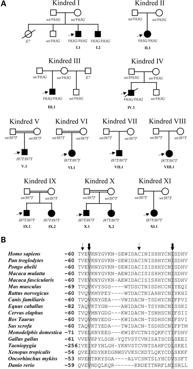
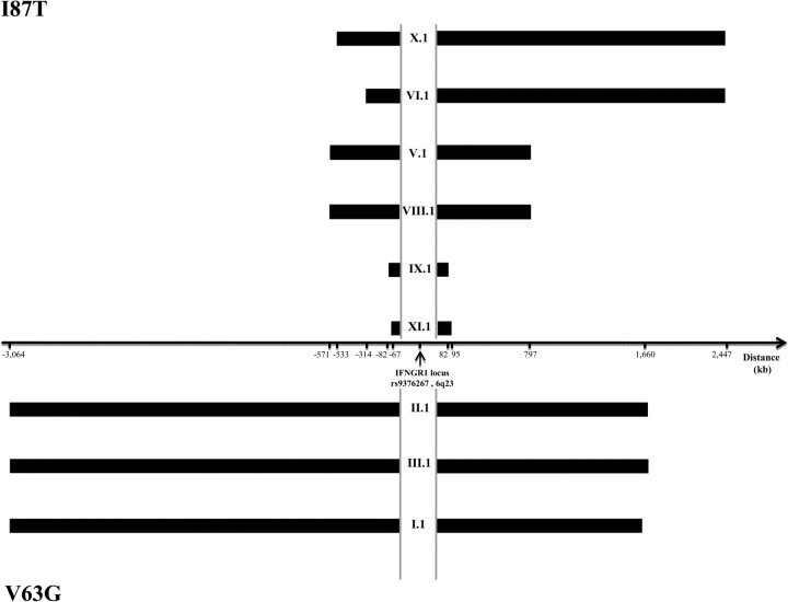
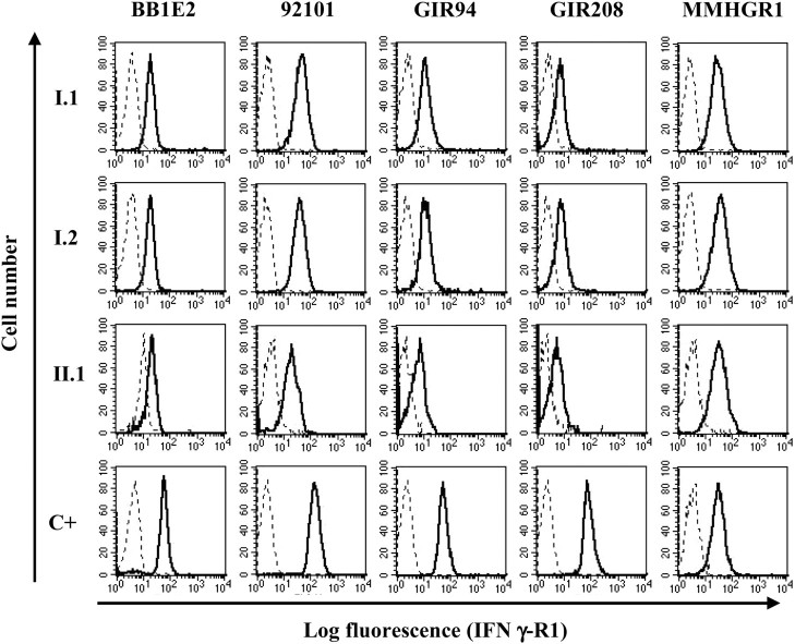
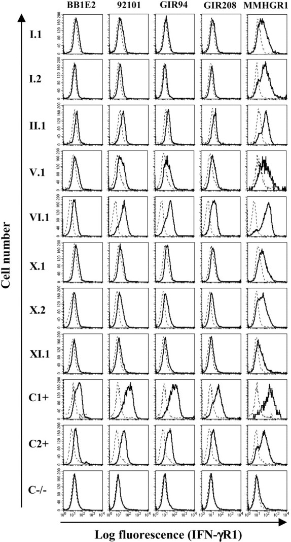
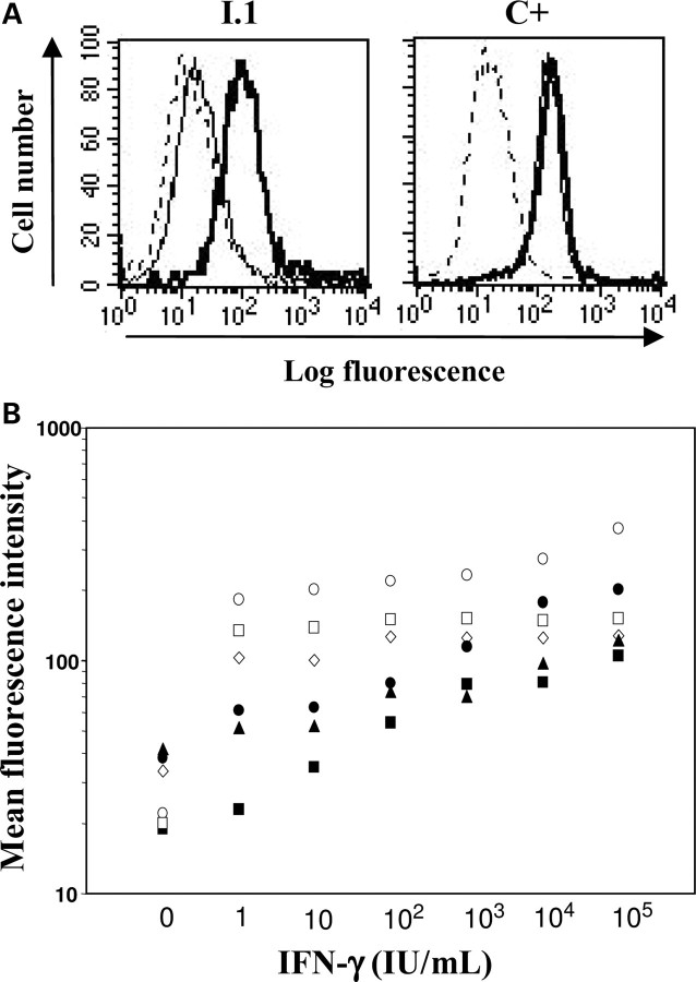
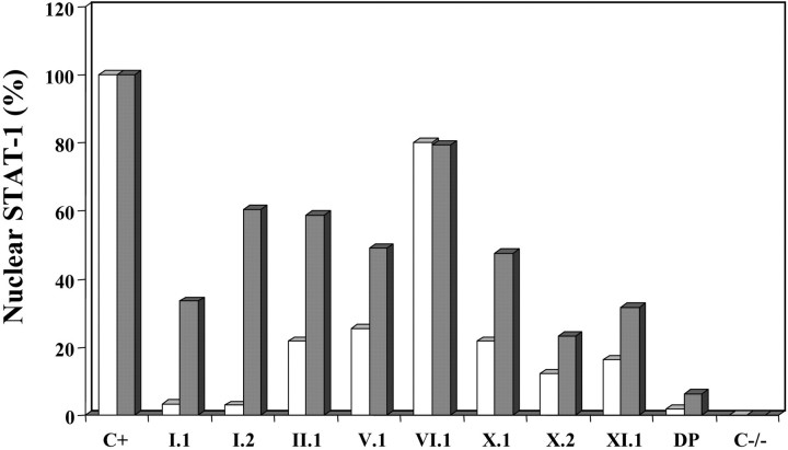
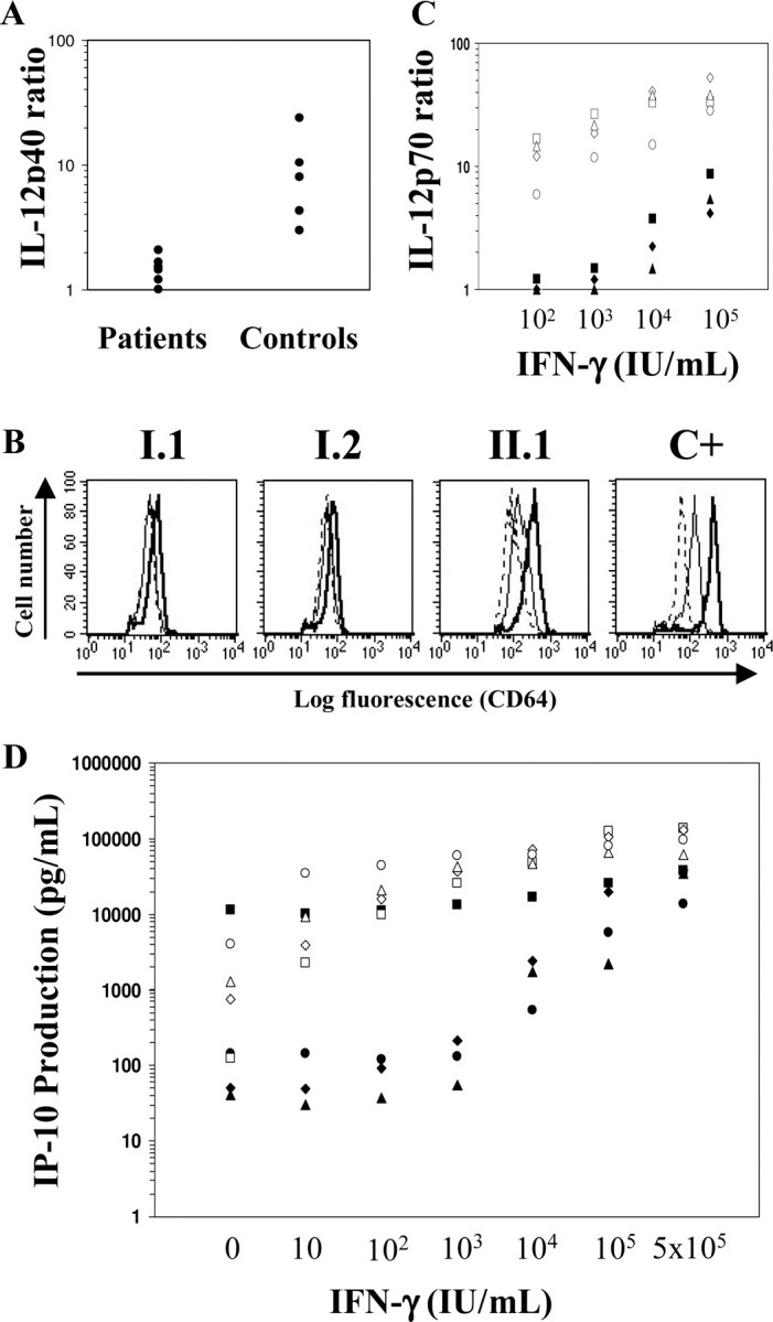
We report a series of 14 patients from 11 kindreds with recessive partial (RP)-interferon (IFN)-γR1 deficiency. The I87T mutation was found in nine homozygous patients from Chile, Portugal and Poland, and the V63G mutation was found in five homozygous patients from the Canary Islands. Founder effects accounted for the recurrence of both mutations. The most recent common ancestors of the patients with the I87T and V63G mutations probably lived 1600 (875-2950) and 500 (200-1275) years ago, respectively. The two alleles confer phenotypes that are similar but differ in terms of IFN-γR1 levels and residual response to IFN-γ. The patients suffered from bacillus Calmette-Guérin-osis (n= 6), environmental mycobacteriosis (n= 6) or tuberculosis (n= 1). One patient did not suffer from mycobacterial infections but had disseminated salmonellosis, which was also present in two other patients. Age at onset of the first environmental mycobacterial disease differed widely between patients, with a mean value of 11.25 ± 9.13 years. Thirteen patients survived until the age of 14.82 ± 11.2 years, and one patient died at the age of 7 years, 9 days after the diagnosis of long-term Mycobacterium avium infection and the initiation of antimycobacterial treatment. Up to 10 patients are currently free of infection with no prophylaxis. The clinical heterogeneity of the 14 patients was not clearly related to either IFNGR1 genotype or the resulting cellular phenotype. RP-IFN-γR1 deficiency is, thus, more common than initially thought and should be considered in both children and adults with mild or severe mycobacterial diseases.


Figures

Figure 1.
Figure 1.
Missense mutations in the gene encoding the IFN-γR1 chain identified in the patients and their relatives. (A) Pedigrees of the 11 unrelated families of the IFN-γR1-deficient patients. The patients are indicated by a black square or circle. The IFNGR1 genotypes (V63G and I87T: the V63G and I87T mutants, respectively; wt, wild-type; E?, unknown) of all family members are indicated. Roman numerals indicate the kindred and Arabic numerals indicate the patients in each kindred. The index patients are indicated by an arrow. (B) Alignment of the extracellular portion of the human IFN-γR1 molecule containing residues 63 and 87 and the corresponding regions in 17 other species. The mutated residues are indicated in gray and by a thick arrow. Residues 61 and 77, found to be mutated (C77Y, C77F, V61Q) in patients with RC-IFN-γR1 deficiency, are indicated by a thin arrow. The alignment was edited manually with Bioedit software (51).
Figure 2.
Figure 2.
Haplotype sharing in the IFNGR1 region, in patients with the I87T and V63G mutations. Long continuous stretches of homozygosity were observed around the gene, consistent with its recessive mode of inheritance. Haplotypes were thus unambiguously derived from genotypes. Perfect haplotype matches are shown after the exclusion of no-call SNPs.
Figure 3.
Figure 3.
Detectable IFN-γR1 molecules on the surface of monocytes of the IFNGR1-V63G-homozygous patients. Whole blood from three V63G-homozygous patients and from one healthy control (C+) was stained with five IFN-γR1-specific mAbs (solid lines) and isotypic control antibodies (dashed line). The specific binding observed in C+ is representative of that observed in monocytes from four healthy controls.
Figure 4.
Figure 4.
IFN-γR1 expression on EBV-B cells from patients homozygous for the V63G and I87T mutations. EBV-B cells from three V63G-homozygous patients (I.1, I.2 and II.1), five I87T-homozygous patients (V.1, VI.1, X.1, X.2 and XI.1), two healthy controls (positive control, C1+ and C2+) and a patient with complete IFN-γR1 deficiency and no expression of IFN-γR1 described in a previous study (negative control, C−/−) were stained with five IFN-γR1-specific mAbs (solid lines) and isotypic control primary antibodies (dashed lines).
Figure 5.
Figure 5.
Cells from IFNGR1-V63G-homozygous patients show impaired but detectable IFN-γ binding. (A) Representative experiment showing the binding of anti-IFN-γ antibody to monocytes from patient I.1 and a healthy control (C+) when cells were incubated with medium alone (dashed line), 1IU IFN-γ/ml (thin line) and 105 IU IFN-γ/ml (thick line). (B) Data are the MFI of anti-IFN-γ antibody binding to monocytes from patients I.1, I.2 and III.1 (closed symbols) and three healthy controls (open symbols). Each symbol represents the value for an individual patient or healthy control.
Figure 6.
Figure 6.
High concentrations of IFN-γ induce STAT-1 phosphorylation in monocytes from IFNGR1-V63G-homozygous patients. Data are the MFI of anti-STAT-1-Y701 antibody binding to monocytes from patients I.1, I.2 and II.1 (black closed symbols), two healthy carriers (gray closed symbols) and two healthy controls (open symbols). Each symbol represents the value from an individual patient or healthy control.
Figure 7.
Figure 7.
Translocation of STAT-1 to the nucleus upon stimulation with IFN-γ, in terms of the effect of the I87T and V63G mutations. EBV-B cells from patients homozygous for the V63G (I.1, I.2, II,1) or the I87T (V.1, VI.1, X.1, X.2, XI.1.) mutation, a healthy control (positive control, C+), and two patients with DP-IFN-γR1 deficiency and complete IFN-γR1 deficiency (negative control, C−/−), respectively, reported elsewhere were incubated without and with 103 (white) or 105 (gray) IU IFN-γ/ml for 30 min. The translocation of STAT-1 into the nucleus was detected by ELISA. Values indicate the percentage binding to DNA in nuclear extracts from patients’ EBV-B cells stimulated with various doses of IFN-γ relative to binding to DNA in EBV-B cells from a healthy control.
Figure 8.
Figure 8.
Late events in IFN-γ-induced activation are detectable in cells from patients homozygous for the V63G mutation. (A) Low levels of IL-12p40 production in response to BCG plus IFN-γ. Symbols represent the ratio of IL-12p40 produced in response to BCG plus IFN-γ to IL-12p40 produced in response to BCG alone, in whole-blood cultures from three V63G-homozygous patients, three I87T-homozygous patients and six healthy controls. (B) Induction of CD64 expression on monocytes following stimulation with IFN-γ. Histograms show the binding of anti-CD64 antibody to whole-blood monocytes from patients I.1, I.2, and II.1 and a healthy control (C+) left unstimulated (dashed line) or stimulated with 10 IU/ml (thin line) or 104 IU/ml (thick line) IFN-γ. The results are representative of two independent experiments for each patient and healthy control. (C) IL-12p70 production in vitro in response to various concentrations of IFN-γ. The data shown are the ratio of IL-12p70 produced in response to LPS plus various concentrations of IFN-γ to IL-12p70 produced in response to LPS alone. Each symbol represents the value from an individual V63G-homozygous patient (closed symbols) or healthy control (open symbols). (D) In vitro IP-10 production in response to various concentrations of IFN-γ.  Each symbol represents the value from an individual V63G-homozygous patient (closed symbols, four patients) or healthy control (open symbols, four healthy controls).

References

    1. Casanova J.L., Abel L. Primary immunodeficiencies: a field in its infancy. Science. 2007;317:617–619. doi:10.1126/science.1142963. - DOI - PubMed
    1. Casanova J.L., Abel L. Genetic dissection of immunity to mycobacteria: the human model. Annu. Rev. Immunol. 2002;20:581–620. doi:10.1146/annurev.immunol.20.081501.125851. - DOI - PubMed
    1. Rosenzweig S.D., Holland S.M. Defects in the interferon-gamma and interleukin-12 pathways. Immunol. Rev. 2005;203:38–47. doi:10.1111/j.0105-2896.2005.00227.x. - DOI - PubMed
    1. Filipe-Santos O., Bustamante J., Chapgier A., Vogt G., de Beaucoudrey L., Feinberg J., Jouanguy E., Boisson-Dupuis S., Fieschi C., Picard C., et al. Inborn errors of IL-12/23- and IFN-gamma-mediated immunity: molecular, cellular, and clinical features. Semin. Immunol. 2006;18:347–361. doi:10.1016/j.smim.2006.07.010. - DOI - PubMed
    1. Alcais A., Fieschi C., Abel L., Casanova J.L. Tuberculosis in children and adults: two distinct genetic diseases. J. Exp. Med. 2005;202:1617–1621. doi:10.1084/jem.20071987. - DOI - PMC - PubMed

Publication types

MeSH terms