. 2011 May;39(10):4166-79.
doi: 10.1093/nar/gkq1363. Epub 2011 Jan 25.

Use of RecA fusion proteins to induce genomic modifications in zebrafish

Affiliations

Use of RecA fusion proteins to induce genomic modifications in zebrafish

Hsin-Kai Liao et al. Nucleic Acids Res. 2011 May.

Abstract

The bacterial recombinase RecA forms a nucleic acid-protein filament on single-stranded (ss) DNA during the repair of double-strand breaks (DSBs) that efficiently undergoes a homology search and engages in pairing with the complementary DNA sequence. We utilized the pairing activity of RecA-DNA filaments to tether biochemical activities to specific chromosomal sites. Different filaments with chimeric RecA proteins were tested for the ability to induce loss of heterozygosity at the golden locus in zebrafish after injection at the one-cell stage. A fusion protein between RecA containing a nuclear localization signal (NLS) and the DNA-binding domain of Gal4 (NLS-RecA-Gal4) displayed the most activity. Our results demonstrate that complementary ssDNA filaments as short as 60 nucleotides coated with NLS-RecA-Gal4 protein are able to cause loss of heterozygosity in ∼3% of the injected embryos. We demonstrate that lesions in ∼9% of the F0 zebrafish are transmitted to subsequent generations as large chromosomal deletions. Co-injection of linear DNA with the NLS-RecA-Gal4 DNA filaments promotes the insertion of the DNA into targeted genomic locations. Our data support a model whereby NLS-RecA-Gal4 DNA filaments bind to complementary target sites on chromatin and stall DNA replication forks, resulting in a DNA DSB.


Figures

Figure 1.
Figure 1.
(A) RecA fusion proteins used in this study. (B) Injection of complementary ssDNA-NLS-RecA-Gal4 filaments leads to LOH at the gol locus, resulting in mosaic eye pigmentation. Genotype of the embryos used for injection is shown. (C) An example of LOH at the gol locus. Dorsal views of eyes at 3 dpf showing wild-type pigmentation patterns in a non-injected embryo (upper panel) and mosaic pigmentation patterns (bottom panel) after injection of complementary ssgol-NLS-RecA-Gal4 targeting filaments. (D) DNA complementary to either exons4/5 or exon6 of gol was used for making gol RecA filaments. Golm1381 (m, mutant; 1381, 1381 bp) and golex6m-60 (ex6, exon 6; m, mutant; 60, 60 bp) DNAs carry mutations resulting in a stop codon (asterisk). Golex6-60-1(ex6, exon 6; 60, 60 bp) and golex6-60-2 (ex6, exon 6; 60, 60 bp) oligonucleotides do not have mutations.
Figure 2.
Figure 2.
Expression of an EGFP gene trap is consistent with site-specific integration into the gol, flh and prom1a loci. Single-stranded NLS-RecA-Gal4 filaments complementary to regions of the gol (A), flh (B) and prom1a (C) genes were co-injected with the EGFP gene trap cassettes. EGFP expression consistent with targeting gene expression was observed in 5–19% of the injected embryos (Table 2). For the gol gene, expression was observed in the retina at 3 dpf (white arrows, A), for the flh gene expression was detected in the notochord at 14 hpf (arrows, B; arrowheads mark the notochord boundary) and for the prom1a gene expression was detected in the dorsal diencephalon at 30 hpf (arrows, C).
Figure 3.
Figure 3.
Complementary ss gol-NLS-RecA-Gal4 filaments direct site-specific insertion of a gene trap into the gol locus. (A) Two regions of the gol gene corresponding to exons 4 through 5 (gol-270, 270 bp) and exon 6 (gol-300) were amplified, denatured and coated with NLS-RecA-Gal4 protein to make complementary ss-gol-NLS-RecA-Gal4 filaments. Filaments were injected with the EGFP gene trap that contains a SA followed by EGFP in three reading frames and a poly adenylation signal (pA). (B) PCR amplification of junction fragments between the EGFP gene trap and the endogenous gol locus was observed with DNA isolated from individual embryos. Only primers proximal to the region complementary to the filament yielded amplification products that could be verified by sequencing (asterisks). (C) Junction fragments allow insertions to be mapped to the gol locus. The gol-270 filaments (black bar) resulted in insertion of the EGFP gene trap 5′ and 3′ to the region complementary to the filaments (black triangles). In some cases, identical junction fragments were observed from different embryos (triangles marked 4×). The triangle marked 4x corresponds to the 3′-end of the gol-270 filament. The gol-300 filament (clear bar) also promoted insertion near regions 5′ to the complementary sequence in the gol gene (clear triangles).
Figure 4.
Figure 4.
Targeted mutations of the gol locus using gol-NLS-RecA-Gal4 filaments were transmitted through the zebrafish germline. (A and B). A testcross between a F0 zebrafish injected with gol-NLS-RecA-Gal4 filaments and a golb1 homozygote produces offspring that fail to complement the golb1 allele (B) and siblings with normal pigment (A).
Figure 5.
Figure 5.
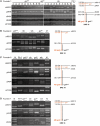
Mapping of new gol alleles from Founders 1, 4, 6 and 7 with z-markers reveals deletions at the gol locus. Founders were crossed with golb1 homozygotes and DNA isolated from offspring displaying the golb1 phenotype and from siblings (sib) with wild-type pigmentation. Parental lines (P0 and golb1) or the wild-type strain used for injection (WIK) is shown for comparison. DNA was amplified with primer pairs to identify SSLPs that were polymorphic. (A) Offspring from Founder 1 that fail to complement the golb1 mutation have one allele for markers SSLPs z46013, golin8, z9404 and z8488, but are heterozygous for marker z21330. This indicates a deletion of 32.5–38.5 CM, with a proximal breakpoint boundary that maps between z8488 and z21330. (B) In offspring from Founder 4 the only SSLP alleles present were from the golb1 parent, indicating the deletion is >38.5 CM. (C) In the offspring from Founder 6 that failed to complement the golb1 mutation, a single allele was present for marker z9404 but heterozygous for marker z21330, indicating a deletion of at least 7 CM. (D) Founder 7 produced offspring with a deletion that contained only marker z9404, indicating a deletion of at least 7 CM.
Figure 6.
Figure 6.
A model for gene targeting using complementary ss DNA-NLS-RecA-Gal4. Both sense and antisense single-strand (ss-s and ss-a)-NLS-RecA-Gal4 filaments are co-injected into zebrafish embryos. The RecA homology search activity can guide the filaments to the targeted region. The cssDNA-NLS-RecA-Gal4 filaments can undergo homologous pairing and strand invasion. This causes the formation of D-loops that are stabilized by the Gal4 dimerization domains between the complementary filaments on the target chromosome. This compact DNA joint molecule is assumed to block replication fork progression, leading to a DNA DSB.

References

    1. McGrew DA, Knight KL. Molecular design and functional organization of the RecA protein. Crit. Rev. Biochem. Mol. Biol. 2003;38:385–432. - PubMed
    1. Radding CM, Shibata T, DasGupta C, Cunningham RP, Osber L. Kinetics and topology of homologous pairing promoted by Escherichia coli recA-gene protein. Cold Spring Harb. Symp. Quant. Biol. 1981;45(Pt 1):385–390. - PubMed
    1. Seitz EM, Brockman JP, Sandler SJ, Clark AJ, Kowalczykowski SC. RadA protein is an archaeal RecA protein homolog that catalyzes DNA strand exchange. Genes Dev. 1998;12:1248–1253. - PMC - PubMed
    1. Cox MM. Recombinational DNA repair in bacteria and the RecA protein. Prog. Nucleic Acid Res. Mol. Biol. 1999;63:311–366. - PubMed
    1. Campbell MJ, Davis RW. On the in vivo function of the RecA ATPase. J. Mol. Biol. 1999;286:437–445. - PubMed

Publication types

MeSH terms