. 2011 Feb 8;108(6):2228-33.
doi: 10.1073/pnas.1015287108. Epub 2011 Jan 25.

Molecular basis for ubiquitin and ISG15 cross-reactivity in viral ovarian tumor domains

Affiliations

Molecular basis for ubiquitin and ISG15 cross-reactivity in viral ovarian tumor domains

Masato Akutsu et al. Proc Natl Acad Sci U S A. .

Abstract

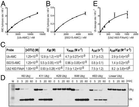
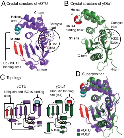
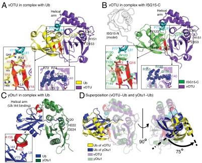
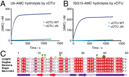
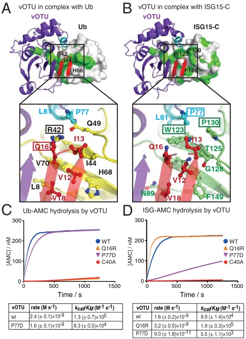
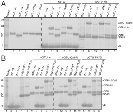
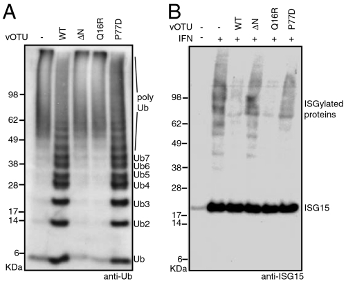
Crimean Congo hemorrhagic fever virus (CCHFV) is a deadly human pathogen that evades innate immune responses by efficiently interfering with antiviral signaling pathways mediated by NF-κB, IRF3, and IFNα/β. These pathways rely on protein ubiquitination for their activation, and one outcome is the modification of proteins with the ubiquitin (Ub)-like modifier interferon-stimulated gene (ISG)15. CCHFV and related viruses encode a deubiquitinase (DUB) of the ovarian tumor (OTU) family, which unlike eukaryotic OTU DUBs also targets ISG15 modifications. Here we characterized the viral OTU domain of CCHFV (vOTU) biochemically and structurally, revealing that it hydrolyzes four out of six tested Ub linkages, but lacks activity against linear and K29-linked Ub chains. vOTU cleaved Ub and ISG15 with similar kinetics, and we were able to understand vOTU cross-reactivity at the molecular level from crystal structures of vOTU in complex with Ub and ISG15. An N-terminal extension in vOTU not present in eukaryotic OTU binds to the hydrophobic Ile44 patch of Ub, which results in a dramatically different Ub orientation compared to a eukaryotic OTU-Ub complex. The C-terminal Ub-like fold of ISG15 (ISG15-C) adopts an equivalent binding orientation. Interestingly, ISG15-C contains an additional second hydrophobic surface that is specifically contacted by vOTU. These subtle differences in Ub/ISG15 binding allowed the design of vOTU variants specific for either Ub or ISG15, which will be useful tools to understand the relative contribution of ubiquitination vs. ISGylation in viral infection. Furthermore, the crystal structures will allow structure-based design of antiviral agents targeting this enzyme.


Conflict of interest statement

Conflict of interest statement: D.K. is a consultant for Ubiquigent, Inc.

Figures

Fig. 1.
Fig. 1.
Cross-reactivity and linkage specificity of CCHFV vOTU. (A) Michaelis–Menten kinetics for vOTU determined by quantitative Ub-AMC measurements. Curve-fitting was performed with GraphPad Prism. (B) Michaelis–Menten kinetics determined with ISG15-AMC. (C) Table summarizing the kinetic parameters for vOTU. (D) Qualitative linkage-specificity analysis of vOTU against diUb of different linkages was performed according to ref. . vOTU (2.5 ng) was incubated with 1.5 μg diUb in 30 μL volume at 37 °C. 5 μL samples were taken at indicated time points, resolved on a 4–12% SDS-PAGE gel and silver stained. (E) Michaelis–Menten kinetics derived from a fluorescence anisotropy assay against FlAsH-tagged K63-linked diUb.
Fig. 2.
Fig. 2.
Structural features of the CCHFV OTU domain. (A) Structure of the vOTU domain in cartoon representation. Secondary structure elements facilitating Ub/ISG15 binding are highlighted in orange/green. Catalytic site residues are shown in ball-and-stick representation with residues labeled. All structure figures were prepared with PyMol (www.pymol.org). (B) Crystal structure of yOtu1 [green; PDB ID 3by4 (23)]. The Ub contacting helix in the helical arm is coloured orange and catalytic site residues are shown. (C) Topology diagrams of vOTU and yOtu1 colored as in A/B. Yellow stars indicate the relative position of catalytic site residues, and Ub/ISG15 interacting regions are indicated. Topology diagrams were prepared with TopDraw. (D) Superposition of vOTU (colored as in A) and yOtu1 (colored as in C).
Fig. 3.
Fig. 3.
Structures of vOTU in complex with Ub and ISG15-C. (A) Structure of vOTU in complex with Ub. The large picture shows the complex, with the OTU domain labeled as in Fig. 2A, and Ub in yellow. Residues mediating the interactions are shown in ball-and-stick representation and labeled. Two insets show the molecular detail of Ub binding to the N-terminal β-sheet (Left) and weighted 2|Fo|-|Fc| electron density contoured at 1σ, for the Ub C-terminus linked to the catalytic site Cys residue (Right). (B) Structure of vOTU in complex with ISG15-C (in green), images according to (A). The N-terminal Ubl-fold of ISG15 is modeled (outlined in black) according to the crystal structure of full-length ISG15 [PDB ID 1z2m (12)]. (C) Structure of yOtu1 (green, with an orange Ub-binding helix) in complex with Ub (blue) [PDB ID 3by4 (23)]. The inset shows the interaction of the Ub-binding helix with the Ub hydrophobic patch. (D) Superposition of vOTU–Ub and yOtul–Ub complexes shown in two orientations. Proteins are colored as in (A) and (D), and OTU domains are shown as semitransparent cartoon. The 75° rotation of Ub along the long helix of Ub is indicated in the right image.
Fig. 4.
Fig. 4.
The vOTU N-terminus is essential and conserved. (A) Ub-AMC assay of vOTU and vOTUΔN. (B) ISG15-AMC assay of vOTU and vOTUΔN. (C) Sequence alignment of the N-terminus in nairoviruses CCHFV (UniProt Accession number: Q6TQR6), Dugbe virus (Q66431), Hazara virus (A6XA53), Kupe virus (B8PWH5), and Nairobi sheep disease virus (D0PRM9). Alignments generated by the T-COFFEE server (http://www.ch.embnet.org/software/TCoffee.html) using regions corresponding to residues 1–183 (Fig. S3) of vOTU were visualized by ESPript 2.2 (http://espript.ibcp.fr/ESPript/ESPript/). Secondary structure elements are shown below the alignment. Residues contacting the Ub/ISG15 hydrophobic patch are indicated by orange or green dots, respectively, and a yellow star indicates the catalytic Cys residue.
Fig. 5.
Fig. 5.
Structural differences between Ub and ISG15 allow switching of vOTU specificity. (A) The top image shows Ub (under a surface with hydrophobic residues colored green) bound to vOTU centered on the N-terminal extension. Ile44 of Ub is indicated on the surface. The image below is a close-up of the interface between Ub (yellow) and vOTU, highlighting the interaction between vOTU Gln16 and Ub Arg42. (B) vOTU in complex with ISG15-C (green) depicted as in (A). Residues Trp123, Pro130, and Phe149 are indicated on the surface of ISG15-C. The image below centers on the vOTU Pro77 interaction with Pro130 and Trp123 of ISG15. (C) Ub-AMC assays for vOTU and mutants. (D) ISG15-AMC assays for vOTU and mutants.
Fig. 6.
Fig. 6.
Analysis of vOTU cross-reactivity using suicide probes. Ub and ISG15 thioesters were converted with 2-chloroethylamine hydrochloride to suicide inhibitors according to ref. , and tested against vOTU and vOTU mutants. Reactions were stopped after 5 min, resolved on SDS-PAGE gels and Coomassie stained. (A) Reaction of vOTU and mutants with Ub and ISG15 suicide probes. (B) Reaction of vOTU, vOTU Q16R, and vOTU P77D with Ub, Ub R42A, ISG15, and ISG15 W123R mutant suicide probes. WT; wild-type vOTU.
Fig. 7.
Fig. 7.
Verification of vOTU mutant specificity against in vivo substrates. (A) PolyUb UBE2S was incubated with wild-type and mutant vOTU and ubiquitinated species were detected by Western blotting with an anti-Ub antibody (Millipore). (B) HeLa cells were stimulated with IFNβ according to ref. , and total cell lysates were incubated with vOTU and mutants. ISG15 was detected by Western blotting with an anti-ISG15 antibody (Santa Cruz). WT; wild-type vOTU.

References

    1. Akira S, Uematsu S, Takeuchi O. Pathogen recognition and innate immunity. Cell. 2006;124:783–801. - PubMed
    1. O’Neill LA, Bowie AG. Sensing and signaling in antiviral innate immunity. Curr Biol. 2010;20:R328–333. - PubMed
    1. Sadler AJ, Williams BRG. Interferon-inducible antiviral effectors. Nat Rev Immunol. 2008;8:559–568. - PMC - PubMed
    1. Komander D. The emerging complexity of protein ubiquitination. Biochem Soc Trans. 2009;37:937–953. - PubMed
    1. Zeng W, et al. Reconstitution of the RIG-I pathway reveals a signaling role of unanchored polyubiquitin chains in innate immunity. Cell. 2010;141:315–330. - PMC - PubMed

Publication types

MeSH terms