. 2011 Mar 18;286(11):8924-32.
doi: 10.1074/jbc.M110.180638. Epub 2011 Jan 25.

Naturally secreted amyloid-beta increases mammalian target of rapamycin (mTOR) activity via a PRAS40-mediated mechanism

Affiliations

Naturally secreted amyloid-beta increases mammalian target of rapamycin (mTOR) activity via a PRAS40-mediated mechanism

Antonella Caccamo et al. J Biol Chem. .

Abstract

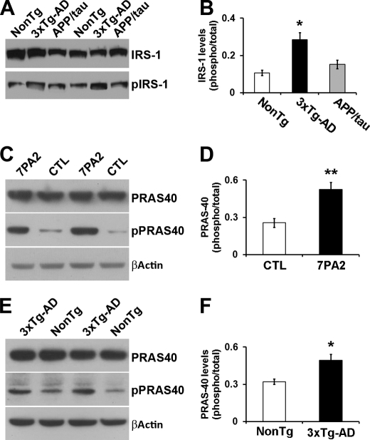
Reducing the mammalian target of rapamycin (mTOR) activity increases lifespan and health span in a variety of organisms. Alterations in protein homeostasis and mTOR activity and signaling have been reported in several neurodegenerative disorders, including Alzheimer disease (AD); however, the causes of such deregulations remain elusive. Here, we show that mTOR activity and signaling are increased in cell lines stably transfected with mutant amyloid precursor protein (APP) and in brains of 3xTg-AD mice, an animal model of AD. In addition, we show that in the 3xTg-AD mice, mTOR activity can be reduced to wild type levels by genetically preventing Aβ accumulation. Similarly, intrahippocampal injections of an anti-Aβ antibody reduced Aβ levels and normalized mTOR activity, indicating that high Aβ levels are necessary for mTOR hyperactivity in 3xTg-AD mice. We also show that the intrahippocampal injection of naturally secreted Aβ is sufficient to increase mTOR signaling in the brains of wild type mice. The mechanism behind the Aβ-induced mTOR hyperactivity is mediated by the proline-rich Akt substrate 40 (PRAS40) as we show that the activation of PRAS40 plays a key role in the Aβ-induced mTOR hyperactivity. Taken together, our data show that Aβ accumulation, which has been suggested to be the culprit of AD pathogenesis, causes mTOR hyperactivity by regulating PRAS40 phosphorylation. These data further indicate that the mTOR pathway is one of the pathways by which Aβ exerts its toxicity and further support the idea that reducing mTOR signaling in AD may be a valid therapeutic approach.


Figures

FIGURE 1.
FIGURE 1.
mTOR signaling and activity are increased in 3xTg-AD mice. A, representative Western blots of protein extracted from the brains of 2-month-old 3xTg-AD and NonTg mice (n = 6/genotype) and probed with the indicated antibodies. B and C, quantitative analyses of the blots show that the p70S6K phosphorylation at Thr-389 (indicated as the ratio of total over phosphorylated levels) and 4E-BP1 phosphorylation at Ser-65 (indicated as the ratio of total over phosphorylated levels) were not significantly different between 3xTg-AD and NonTg mice. D, representative Western blots of protein extracted from the brains of 6-month-old 3xTg-AD and NonTg mice (n = 6/genotype) and probed with the indicated antibodies. E and F, quantitative analyses of the blots show that the p70S6K phosphorylation at Thr-389 (indicated as the ratio of total over phosphorylated levels) and 4E-BP1 phosphorylation at Ser-65 (indicated as the ratio of total over phosphorylated levels) were significantly higher in the brains of the 3xTg-AD mice when compared with NonTg mice (n = 6/genotype). G, representative Western blots of protein extracted from the brains of 12-month-old 3xTg-AD and NonTg mice (n = 6/genotype) and probed with the indicated antibodies. H and I, quantitative analyses of the blots show that the p70S6K phosphorylation at Thr-389 (indicated as the ratio of total over phosphorylated levels) and 4E-BP1 phosphorylation at Ser-65 (indicated as the ratio of total over phosphorylated levels) were significantly higher in the brains of the 3xTg-AD mice when compared with NonTg mice (n = 6/genotype). J, mTOR enzymatic activity was significantly increased in the brains of 6- and 12-month old 3xTg-AD mice when compared with age- and gender-matched NonTg mice. In contrast, no differences were detected at 2 months of age (n = 6/genotype/time point). Data are presented as means ± S.E. *, p < 0.05. **, p < 0.01.
FIGURE 2.
FIGURE 2.
Genetically preventing Aβ accumulation in 3xTg-AD mice reduces mTOR activity. A and B, representative microphotographs showing CA1 pyramidal neurons of 6-month-old 3xTg-AD and APP/Tau mice (n = 6/genotype). Sections were stained with an anti-Aβ antibody. C, representative Western blots of protein extracted from the brains of 6-month-old 3xTg-AD and APP/Tau mice (n = 6/genotype) and probed with the indicated antibodies. D, quantitative analyses of the blots show that p70S6K phosphorylation at Thr-389 (indicated as the ratio of total over phosphorylated levels) was significantly reduced in the APP/Tau mice when compared with 3xTg-AD mice. E, mTOR enzymatic activity was significantly reduced in the brains of 6-month-old APP/Tau mice when compared with age-matched 3xTg-AD mice. Notably, mTOR activity was not statistically significant between NonTg and APP/Tau mice, suggesting that high Aβ levels are necessary to induce mTOR hyperactivity in the 3xTg-AD mice. (n = 6/genotype). Data are presented as means ± S.E. *, p < 0.05.
FIGURE 3.
FIGURE 3.
Lowering Aβ levels reduces mTOR activity. 6E10 was stereotaxically injected into the left hippocampi of 6-month-old mice (n = 6). The right uninjected hippocampi were used as internal controls. A, sandwich ELISA measurements show that Aβ levels were significantly reduced in the ipsilateral (Ipsi) hippocampi (receiving 6E10) when compared with the contralateral (Contra) uninjected hippocampi (n = 6). B, representative Western blots of protein extracted from the left hippocampi of 6-month-old 3xTg-AD injected with 6E10 (ipsilateral) and the right hippocampi used as internal control (contralateral; n = 6) and probed with the indicated antibodies. C, quantitative analyses of the blots show that reducing Aβ levels with 6E10 lowered p70S6K phosphorylation at Thr-389 (indicated as the ratio of total over phosphorylated levels) in the ipsilateral hippocampi (receiving the anti-Aβ antibody). No changes were detected in the contralateral hippocampi. D, mTOR enzymatic activity was significantly reduced in the hippocampi of 6-month-old 3xTg-AD mice receiving 6E10 when compared with contralateral uninjected hippocampi. (n = 6/group). Data are presented as means ± S.E. *, p < 0.05. **, p < 0.01.
FIGURE 4.
FIGURE 4.
Soluble Aβ is sufficient to increase mTOR activity. A, representative Western blots of protein extracted from the hippocampi of 2-month-old NonTg mice stereotaxically injected with concentrated CM from CHO or 7PA2 cells (n = 6/group). Six additional mice were injected with 7PA2 CM that were precleared of Aβ with 6E10. B, quantitative analyses of the blots indicate that injection of CHO CM or precleared 7PA2 CM did not significantly change the phosphorylation of p70S6K at Thr-389 (indicated as the ratio of total over phosphorylated levels). In contrast, injections of 7PA2 CM (highly enriched in soluble Aβ oligomers) significantly increased p70S6K phosphorylation at Thr-389. C, mTOR enzymatic activity was significantly increased in the hippocampi of 2-month-old NonTg mice injected with 7PA2 CM when compared with the hippocampi of mice injected with CHO CM or precleared 7PA2 CM (n = 6/group). Data are presented as means ± S.E. *, p < 0.05.
FIGURE 5.
FIGURE 5.
Phosphorylation of PRAS40 is significantly increased in the hippocampi of 3xTg-AD mice. A, representative Western blots of protein extracted from 6-month-old NonTg, 3xTg-AD, and APP/Tau mice and probed with the indicated antibodies (n = 6/genotype). B, quantitative analysis of the blots indicates that the ratio pIRS1/IRS1 was significantly increased in 3xTg-AD mice when compared with NonTg mice and APP/Tau mice. No difference was detected between the NonTg and APP/Tau mice. C, representative Western blots of protein extracted from 7PA2 and control (CTL) CHO cells and probed with the indicated antibodies (n = 6/group). D, quantitative analyses of the blots indicate that phosphorylation of PRAS40 at Thr-246 (indicated as the ratio of total over phosphorylated levels) was significantly increased in the 7PA2 cells when compared with control cells. E, representative Western blots of protein extracted from the hippocampi of 6-month-old 3xTg-AD and NonTg mice and probed with the indicated antibodies (n = 6/group). F, quantitative analyses of the blots indicate that phosphorylation of PRAS40 at Thr-246 (indicated as the ratio of total over phosphorylated levels) was significantly increased in the hippocampi of the 3xTg-AD mice when compared with NonTg mice. Data are presented as means ± S.E. *, p < 0.05. **, p < 0.01.
FIGURE 6.
FIGURE 6.
PRAS40 phosphorylation plays a key role in the Aβ-mediated increase in mTOR activity. A, representative Western blots of protein extracted from the hippocampi of 2-month-old NonTg mice stereotaxically injected with concentrated CM from CHO and 7PA2 cells in the presence or absence of the Akt inhibitor VII, TAT-Akt-in (Akt inh) or the PIM-1 inhibitor 1 (PIM-1 inh) (n = 6/group). B, quantitative analyses of the blots indicate that injection of 7PA2 CM significantly increased PRAS40 phosphorylation (indicated as the ratio of total over phosphorylated levels). However, when 7PA2 CM was injected with the Akt or PIM-1 inhibitors, PRAS40 phosphorylation levels were significantly reduced when compared with baseline levels (mice injected with CHO CM), indicating that the inhibitor was able to block PRAS40 in vivo. C, mTOR enzymatic activity was significantly increased in the hippocampi of 2-month-old NonTg mice injected with 7PA2 CM when compared with the hippocampi of mice injected with CHO CM, 7PA2 CM + Akt inhibitor VII, TAT-Akt-in, and 7PA2 CM + PIM-1 inhibitor 1 (n = 6/group), suggesting that PRAS40 phosphorylation is necessary for the Aβ-induced increase in mTOR activity. D, representative Western blots of protein extracted from the hippocampus of 6-month-old mice and probed with the indicated antibodies. E, quantitative analyses of the blots indicate that the phosphorylation levels of TSC-1 were similar between the three groups of mice. Data are presented as means ± S.E. *, p < 0.05. **, p < 0.01.

References

    1. Querfurth H. W., LaFerla F. M. (2010) N. Engl. J. Med. 362, 329–344 - PubMed
    1. Grundke-Iqbal I., Iqbal K., Tung Y. C., Quinlan M., Wisniewski H. M., Binder L. I. (1986) Proc. Natl. Acad. Sci. U.S.A. 83, 4913–4917 - PMC - PubMed
    1. Ihara Y., Nukina N., Miura R., Ogawara M. (1986) J. Biochem. 99, 1807–1810 - PubMed
    1. Kosik K. S., Joachim C. L., Selkoe D. J. (1986) Proc. Natl. Acad. Sci. U.S.A. 83, 4044–4048 - PMC - PubMed
    1. Glenner G. G., Wong C. W. (1984) Biochem. Biophys. Res. Commun. 120, 885–890 - PubMed

Publication types

MeSH terms

Substances