. 2011 Jan 25;4(157):ra4.
doi: 10.1126/scisignal.2001225.

Genome-wide RNAi screen reveals disease-associated genes that are common to Hedgehog and Wnt signaling

Affiliations

Genome-wide RNAi screen reveals disease-associated genes that are common to Hedgehog and Wnt signaling

Leni S Jacob et al. Sci Signal. .

Abstract

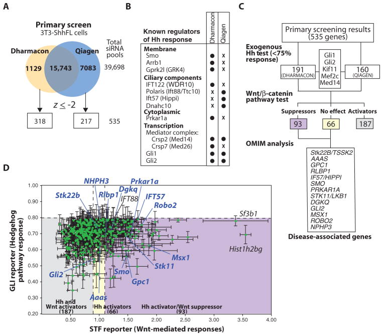
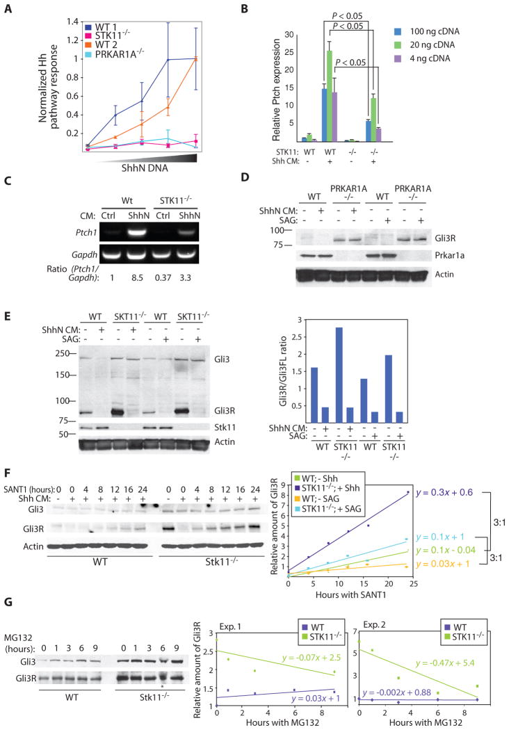
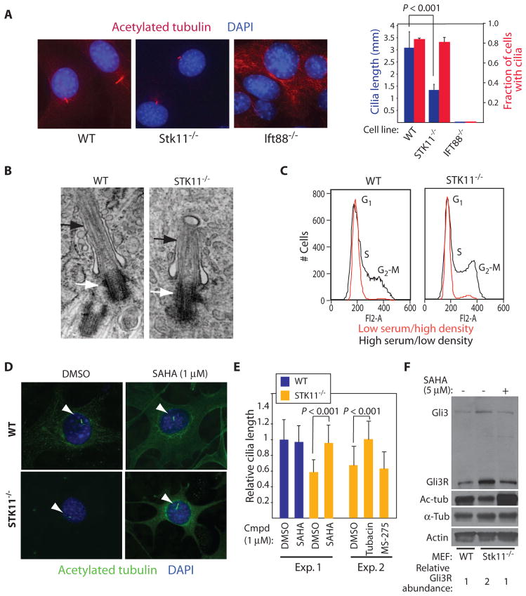
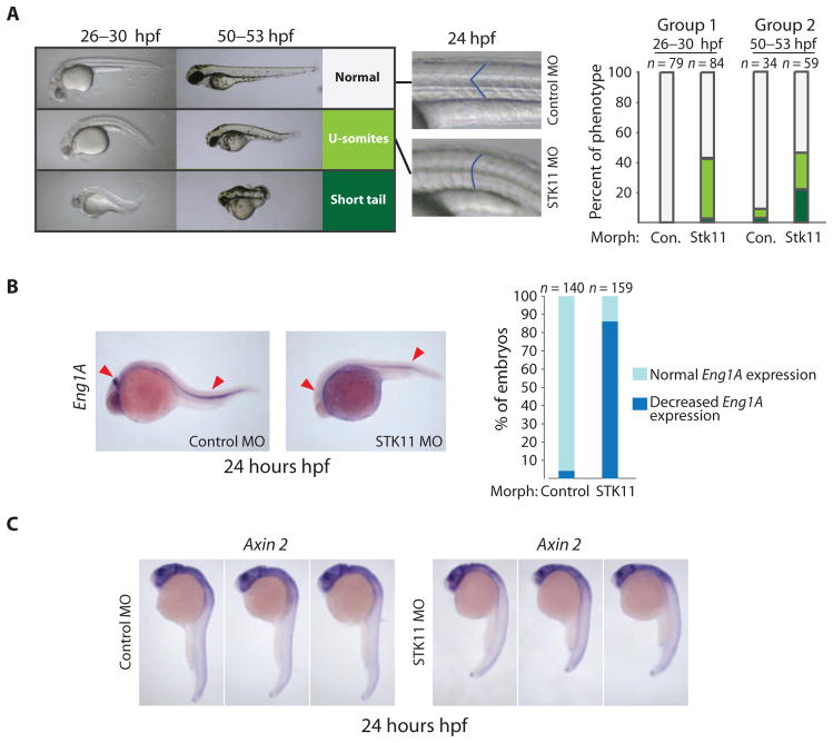
The Hedgehog (Hh) and Wnt signal transduction pathways are master regulators of embryogenesis and tissue renewal and represent anticancer therapeutic targets. Using genome-wide RNA interference screening in murine cultured cells, we established previously unknown associations between these signaling pathways and genes linked to developmental malformations, diseases of premature tissue degeneration, and cancer. We identified functions in both pathways for the multitasking kinase Stk11 (also known as Lkb1), a tumor suppressor implicated in lung and cervical cancers. We found that Stk11 loss resulted in disassembly of the primary cilium, a cellular organizing center for Hh pathway components, thus dampening Hh signaling. Loss of Stk11 also induced aberrant signaling through the Wnt pathway. Chemicals that targeted the Wnt acyltransferase Porcupine or that restored primary cilia length by inhibiting the tubulin deacetylase HDAC6 (histone deacetylase 6) countered deviant pathway activities driven by Stk11 loss. Our study demonstrates that Stk11 is a critical mediator in both the Hh and the Wnt pathways, and our approach provides a platform to support the development of targeted therapeutic strategies.


Conflict of interest statement

Competing interests: L.L., B.C., W.T., and M.E.D. are listed as inventors in a pending patent application associated with the IWR/IWP compounds described in this study.

Figures

Fig. 1
Fig. 1
A genome-scale screen to identify Hh signal transducing genes. (A) siRNA libraries designed by Dharmacon or Qiagen were screened in triplicate using the 3T3-ShhFL cells. (B) Many known Hh pathway components were identified with siRNAs from a single library. (C) Genes identified from the primary screen were retested using NIH 3T3 cells stably harboring the GliBS reporter (ShhLightII cells) in the presence of culture medium containing ShhN (“Exogenous Hh test”). siRNAs that retained their activity in this assay were tested with the STF reporter in NIH 3T3 cells transfected with Wnt3A cDNA to assign function in the Wnt/β-catenin pathway. Genes with no activity or suppressor function in this pathway were cross-referenced with the OMIM database to identify disease-associated genes (Table 1). Multiple names for a single gene are separated by a slash. (D) Graphical summary of gene function in the Hh (y axis) and Wnt/β-catenin (x axis) pathways. Disease-associated genes (blue) are noted along with other genes of interest (black).
Fig. 2
Fig. 2
Loss of Hh pathway response induced by compromised Stk11 or Prkar1a function is associated with increased abundance of GliR. (A) MEFs null for either STK11 or PRKAR1A fail to achieve normal levels of Hh pathway response. MEFs were transfected with the GliBS and control reporters and increasing amounts of ShhN cDNA. Mean and SD are shown. The experiment was performed in triplicate. (B) STK11/ MEFs have dampened transcriptional response to exogenous ShhN. qPCR analysis of Ptch1 expression in wild-type (WT) and STK11/ MEFs in the presence or absence of Shh stimulation. PCR was performed from different concentrations of total cDNA generated from RNA samples, analyzed, and compared to test reproducibility. Ptch1 transcript abundance was normalized to Gapdh (glyceraldehyde-3-phosphate dehydrogenase). Graph shows mean and SD from three samples. (C) RT-PCR–based confirmation of qPCR results. Ptch1 RT-PCR results from WT and STK11/ cells in the presence and absence of Shh-conditioned medium (CM) also show reduced responsiveness of STK11/ cells to Shh. (D) MEFs deficient in Prkar1a exhibit increased formation of Gli3R, even in the presence of Hh signaling mediated by addition of ShhN CM or SAG, as measured with Western blot analysis. Data shown are representative of two experiments. (E) Stk11 regulates Gli3R abundance. STK11/ MEFs exhibit increased abundance of Gli3R that is inhibited by Hh pathway activation. Graph shows quantification of results by densitometry. A representative of three independent experiments is shown. (F) STK11/MEFs exhibit an accelerated rate of Gli3R accumulation. WT or STK11/ MEFs were treated with Shh CM overnight to inhibit Gli3 processing. Shh CM was then removed and replaced with the Smo inhibitor SANT1 to allow processing to proceed. Cells were lysed at indicated time points. Quantification (right) indicates that the rate of Gli3R accumulation is three times faster in the absence of Stk11 as calculated from two independent experiments (one with Shh CM and another with SAG to inhibit Gli3R formation; see fig. S3B). Rate of Gli3R formation was calculated from the slope of each line. Gli3R abundance is calculated relative to abundance of Gli3R in WT cells at 0 hours of SANT1 treatment in the absence of Shh CM or SAG. (G) Gli3R destruction is accelerated rather than slowed in STK11/ MEFs. WT or STK11/ MEFs were treated with the proteasome inhibitor MG132 for various time periods to restrict new formation of Gli3R by proteasome-dependent proteolytic processing. Gli3R abundance was determined by Western blot analysis and quantified by normalizing to Gli3R in WT cells in the absence of MG132. The asterisk indicates result not used for quantification. Data shown are representative of two experiments.
Fig. 3
Fig. 3
Stk11 is required for primary cilia maintenance in embryonic fibroblasts. (A) MEFs were immunostained for acetylated tubulin to detect the primary cilium. Unlike cells lacking the ciliary component IFT88, a rudimentary primary cilium is observed in STK11/ cells. Cells lacking Stk11 do not have a defect in establishing cilia. MEFs of the indicated genotype were scored for the presence of cilia and cilia length by immunofluorescence. Data show the mean and SD of >35 cells per condition. (B) Ultrastructural analysis of the primary cilium in STK11/ cells by transmission electron microscopy. No gross defects in the basal body (white arrows) and axoneme (black arrows) are observed in STK11/cells. Image of STK11/ cilia is representative of four samples. (C) WT and STK11/ MEFs show comparable cell cycle response to serum and cell density. Cells were grown in either high-serum/low-density conditions or low-serum/high-density conditions for 24 hours, stained with propidium iodide, and subjected to FACS (fluorescence-activated cell sorting) analysis. (D) HDAC inhibition restores primary cilium length in STK11/ cells. MEFs treated with the nonselective HDAC inhibitor SAHA were stained for acetylated tubulin. DMSO, dimethyl sulfoxide. (E) HDAC6 inhibition restores primary cilium of STK11/ cells to normal length. Quantification of results in (D) (Exp. 1) and analysis of the specific HDAC6 inhibitor tubacin and the HDAC1 inhibitor MS-275 on cilia length in STK11/ cells (Exp. 2). Fifty cells were counted in each experiment. (F) SAHA treatment of STK11/ MEFs reduces the abundance of Gli3R. Abundance of Gli3R relative to actin was quantified by Western blot analysis and normalized to WT sample. Acetylated tubulin abundance serves as a positive control for SAHA activity. Data are representative of three experiments.
Fig. 4
Fig. 4
Loss of Stk11 engages aberrant Wnt-dependent cellular responses. (A) Phosphorylated Dvl2 is abundant in MEFs lacking Stk11. Data are representative of three experiments. (B) The IWP compound inhibits Porcupine (Porcn), an acyltransferase essential for Wnt protein production. (C) Increased Dvl2 and Dvl3 phosphorylation in STK11/ cells is inhibited by IWP (2.5 μM), indicating that Dvl phosphorylation is dependent on production of Wnt. Results performed from three independent experiments are quantified. The abundance of Dvl1 is increased in STK11/ cells compared to WT cells, and the relative abundance of phosphorylated/unphosphorylated proteins was not quantified. (D) Application of the CK1 inhibitor D4476 (0.2 mM) to STK11/ cells inhibits Dvl2 phosphorylation. Data are representative of three experiments. (E) Loss of Dvl2 inhibits growth of STK11/ cells. MEFs transfected with Dvl2 siRNAs were plated at clonal density, and CellTiter-Glo assay was performed after 6 days. Data show the mean and SD of three samples. (F) Loss of Stk11 is associated with mislocalization of Dvl2. MEFs, transfected with either Dvl2-GFP or Axin1-GFP DNA, were treated with DMSO or IWP. Green, GFP; blue, DAPI (4′,6- diamidino-2-phenylindole). Images were taken at 40× magnification.
Fig. 5
Fig. 5
Zebrafish embryos in which Stk11 is reduced exhibit phenotypes consistent with Hh and Wnt signaling defects. (A) Zebrafish injected with MO against STK11 show U-shaped somites, shortened tails, or no detectable change in body patterning. Phenotypic analysis was performed at two different developmental stages (groups 1 and 2) in separate experiments. The left side shows representative embryos of each phenotype and the right side shows quantification. hpf, hours postfertilization. (B) Loss of Stk11 reduces expression of Engrailed 1A (Eng1A), a target gene induced by Hh signaling. Quantification of embryos with decreased Eng1A expression at the midbrain-hindbrain boundary and muscle pioneers. In situ hybridization with digoxigenin-labeled antisense probes against Eng1a mRNA was performed at 24 hpf embryos. More than 100 animals were scored in each condition. (C) STK11 morphants exhibit little change in Axin2 expression when compared to control animals. In situ hybridization with digoxigenin-labeled antisense probes against Axin2 mRNA was performed at 24 hpf embryos that had been injected with control MO or STK11 MO. Representative embryos from 30 animals analyzed in each group are shown.
Fig. 6
Fig. 6
Loss of Stk11 results in deviant Wnt-mediated responses in cancer cell lines. (A) Cervical carcinoma cells lacking Stk11 exhibit increased abundance of phosphorylated Dvl2 but not necessarily activation of the canonical Wnt/β-catenin pathway. Lysates from cervical carcinoma cell lines or normal endometrial cells (Endo cells) were Western-blotted for Dvl2, Stk11, or c-Myc, the product of a Wnt/β-catenin target gene. Kif3a is a loading control. The asterisk indicates background band. (B) Wnt/β-catenin pathway activity in HeLa cells is sensitive to disruption of Wnt production or inhibition of the canonical Wnt pathway. Endo, HeLa, or SiHA cells transfected with the STF reporter were treated with either IWP or IWR compounds. Data show the mean and SD of three samples. (C) Stk11 disrupts Wnt/β-catenin response in HeLa cells. STF reporter was transfected with or without Wnt3A and Stk11 DNA into HeLa cells. Data show the mean and SD of three samples. (D) Introduction of Stk11 increases the number of HeLa cells with punctate Dvl2 localization. Dvl2-GFP protein in HeLa cells shows a diffuse localization pattern, similar to that observed in STK11/ MEFs. By contrast, the Dvl binding partner Axin1 exhibits a punctate expression pattern (fig. S10). Cells were transfected with Dvl2-GFP DNA with or without Stk11 DNA. n = 50 for each condition. (E) Kinase activity of Stk11 is required for suppression of Dvl2 activity. Expression constructs encoding wild-type or a K78M mutant (kinase-dead, KD) form of Stk11 were transfected into HeLa cells, and effects on Dvl2 phosphorylation were analyzed by Western blotting. Data are representative of three independent experiments. (F) Introduction of Stk11 in HeLa cells inhibits cell growth. Transfected cells were seeded at clonal density. The amount of cellular ATP was determined as a measure of cell number after 5 days. Data show the mean and SD of three samples. (G) Chemically tractable mechanisms underlying aberrant Hh and Wnt pathway responses in STK11-null cells. HDAC6 inhibitors, such as SAHA or tubacin, could restore normal cilia length and the abundance of GliR molecules. Porcn and Axin stabilizers that respectively inhibit Wnt protein production and induce β-catenin destruction could be used to counter the effects of excess Dvl activation.

References

    1. Jiang J, Hui CC. Hedgehog signaling in development and cancer. Dev Cell. 2008;15:801–812. - PMC - PubMed
    1. MacDonald BT, Tamai K, He X. Wnt/β-catenin signaling: Components, mechanisms, and diseases. Dev Cell. 2009;17:9–26. - PMC - PubMed
    1. Barker N, Clevers H. Mining the Wnt pathway for cancer therapeutics. Nat Rev Drug Discov. 2006;5:997–1014. - PubMed
    1. Rubin LL, de Sauvage FJ. Targeting the Hedgehog pathway in cancer. Nat Rev Drug Discov. 2006;5:1026–1033. - PubMed
    1. Jacob L, Lum L. Deconstructing the Hedgehog pathway in development and disease. Science. 2007;318:66–68. - PMC - PubMed

Publication types

MeSH terms

LinkOut - more resources