

Comment
doi: 10.1172/JCI46038.
Epub 2011 Jan 25.
Navigational error in the heart leads to premature ventricular excitation
Affiliations
- PMID: 21266771
- PMCID: PMC3026751
- DOI: 10.1172/JCI46038
Item in Clipboard
Comment
Navigational error in the heart leads to premature ventricular excitation
J Clin Invest.
2011 Feb.
Abstract
In the normal heart, an insulating barrier separates the atria and ventricles. The only way in which electrical impulses can cross this barrier is via the atrioventricular (AV) node, which delays impulse conduction to ensure the forward flow of the blood. However, in some individuals, additional muscular bundles (accessory pathways) allow rapid conduction of electrical impulses from the atria to the ventricles, resulting in premature ventricular excitation and contraction. In this issue of the JCI, two independent research groups demonstrate that erroneous development of the embryonic AV canal, which performs a similar function to that of the adult AV node, is a novel mechanism by which accessory pathways can form.
Figures
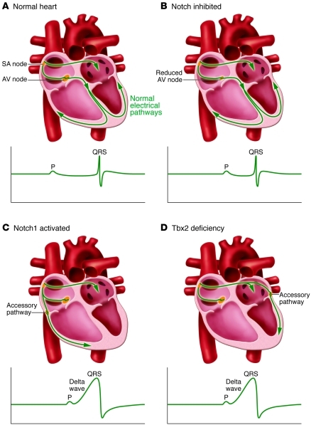
(A) In normal hearts, the electric impulses initiated by pacemaker cells in the sinoatrial (SA) node propagate through the atrial myocardium and trigger its contraction. At the AV node, the impulses are delayed for a period to facilitate alternating contraction of the atrial and ventricular myocardium. After the AV delay, the electrical impulses rapidly travel to the ventricular myocardium via the His-Purkinje system and stimulate the ventricular myocardium. (B) In Notch-inhibited hearts, the AV node is hypoplastic, and the PR interval on the ECG is shortened due to disruption of the AV nodal delay. (C) In Notch1-activated and (D) Tbx2-deficient hearts, accessory pathways are formed as a result of malformation of the AV canal myocardium. The accessory pathways are commonly right-sided in Notch1-activated mice and left-sided in Tbx2-deficient mice. Because of faster conduction through the accessory pathways than through the AV node, the ventricular myocardium is prematurely stimulated (preexcitation). The ECG shows a short PR interval, a slurred upstroke (“delta wave”) of the QRS complex, and a widened QRS complex.
Comment on
-
Defective Tbx2-dependent patterning of the atrioventricular canal myocardium causes accessory pathway formation in mice.J Clin Invest. 2011 Feb;121(2):534-44. doi: 10.1172/JCI44350. Epub 2011 Jan 25. J Clin Invest. 2011. PMID: 21266775 Free PMC article.
-
Notch signaling regulates murine atrioventricular conduction and the formation of accessory pathways.J Clin Invest. 2011 Feb;121(2):525-33. doi: 10.1172/JCI44470. Epub 2011 Jan 25. J Clin Invest. 2011. PMID: 21266778 Free PMC article.
