. 2011 Feb;121(2):534-44.
doi: 10.1172/JCI44350. Epub 2011 Jan 25.

Defective Tbx2-dependent patterning of the atrioventricular canal myocardium causes accessory pathway formation in mice

Affiliations

Defective Tbx2-dependent patterning of the atrioventricular canal myocardium causes accessory pathway formation in mice

Wim T J Aanhaanen et al. J Clin Invest. 2011 Feb.

Abstract

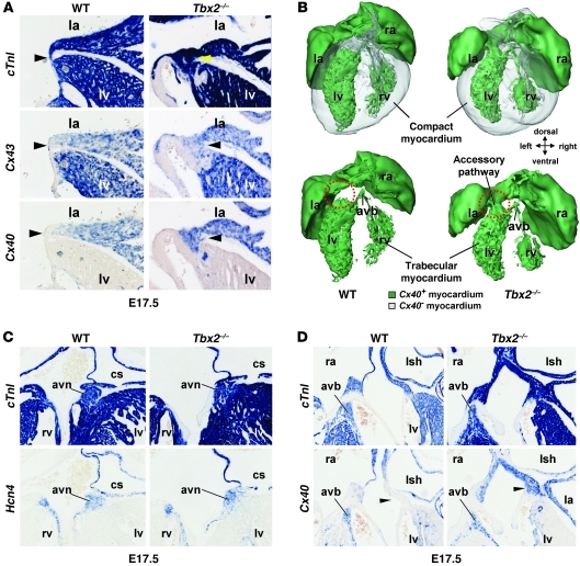
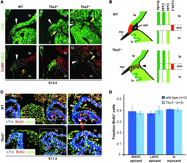
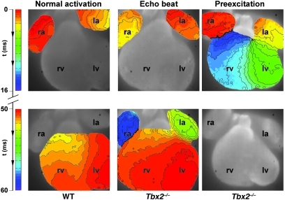
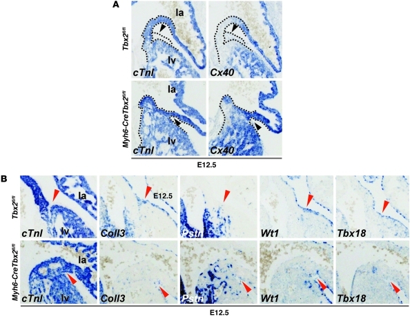
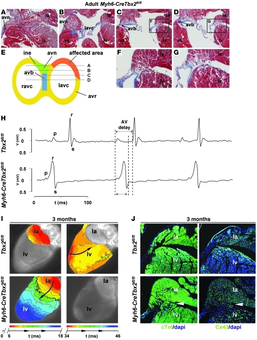
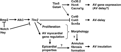
Ventricular preexcitation, a feature of Wolff-Parkinson-White syndrome, is caused by accessory myocardial pathways that bypass the annulus fibrosus. This condition increases the risk of atrioventricular tachycardia and, in the presence of atrial fibrillation, sudden death. The developmental mechanisms underlying accessory pathway formation are poorly understood but are thought to primarily involve malformation of the annulus fibrosus. Before birth, slowly conducting atrioventricular myocardium causes a functional atrioventricular activation delay in the absence of the annulus fibrosus. This myocardium remains present after birth, suggesting that the disturbed development of the atrioventricular canal myocardium may mediate the formation of rapidly conducting accessory pathways. Here we show that myocardium-specific inactivation of T-box 2 (Tbx2), a transcription factor essential for atrioventricular canal patterning, leads to the formation of fast-conducting accessory pathways, malformation of the annulus fibrosus, and ventricular preexcitation in mice. The accessory pathways ectopically express proteins required for fast conduction (connexin-40 [Cx40], Cx43, and sodium channel, voltage-gated, type V, α [Scn5a]). Additional inactivation of Cx30.2, a subunit for gap junctions with low conductance expressed in the atrioventricular canal and unaffected by the loss of Tbx2, did not affect the functionality of the accessory pathways. Our results suggest that malformation of the annulus fibrosus and preexcitation arise from the disturbed development of the atrioventricular myocardium.


Figures

Figure 1
Figure 1. In the left side of the AV canal of Tbx2–/– fetuses, working myocardial genes are ectopically expressed and connect the left atrium with the left ventricle, while the AV node and AV bundle are unaffected.
(A, C, and D) In situ hybridization analyses of serial sections wild-type and Tbx2–/– fetuses at E17.5. (A) In wild-type fetuses, the left AV canal myocardium (arrowheads) does not express Cx40 and Cx43. Tbx2–/– fetuses ectopically expressed Cx40 and Cx43 in the left AV canal (arrowheads). Furthermore, the AV canal in Tbx2–/– fetuses was broader. (B) 3D reconstructions of the heart of wild-type and Tbx2–/– fetuses at E17.5. Green represents Cx40-positive myocardium, and gray represents Cx40-negative myocardium. Only, in Tbx2–/– fetuses, a Cx40-positive myocardial connection formed through the left AV canal (red circles). (C) In the top panels, cTnI reveals the myocardium. The bottom panels show the AV node based on Hcn4 expression and location. The AV node is not affected in Tbx2–/– fetuses. (D) In the top panels, cTnI reveals the myocardium. The bottom panels show the AV bundle based on Cx40 expression and location. The AV bundle is not affected in Tbx2–/– fetuses. la, left atrium; ra, right atrium; avb, AV bundle; avn, AV node; cs, coronary sinus; lsh, left sinus horn. Original magnification, ×10 (A); ×5 (C and D).
Figure 2
Figure 2. In the left side of AV canal of Tbx2–/– fetuses working myocardial proteins are ectopically expressed.
The proliferation rate in the epicardium is not different between wild-type and Tbx2–/– fetuses. (A) Immunohistochemical analyses of serial section of E14.5 wild-type fetuses and 2 Tbx2–/– fetuses (f1, f2). In wild-type fetuses, Cx30.2 is expressed in the AV canal complementary to expression of Cx40. In Tbx2–/– fetuses, Cx40 is expressed ectopically in the AV canal, and Cx30.2 is still expressed in the AV canal myocardium. Notice the variable size and morphology of the aberrant myocardial connection. The white and yellow arrowheads point to the AV canal myocardium. The dashed lines mark the myocardium that connects the left atrium and the left ventricle. (B) Schematic representation of the expression profiles of connexins in the left AV canal myocardium in wild-type and Tbx2–/– fetuses. Note that Cx40 expression withdraws from the compact myocardium; however, Cx43 remains present. Dots represent nonmyocardial cells of the malformed annulus fibrosus. (C) Immunohistochemical analyses of BrdU incorporation in epicardial cells at the left side of the AV canal in a wild-type and Tbx2–/– fetus. The panels on the right are higher magnification images of the areas within the white squares in the panels on the left. The white arrowhead points to the AV myocardium. The red arrowhead points to BrdU+ myocardial cells in the AV myocardium. The yellow arrowhead points to a BrdU+ epicardial cell. (D) A bar graph representing the proliferation rate, based on BrdU incorporation in the myocardium (myocard) of the left ventricle and in the epicardium (epicard) at the right and left side of the AV canal. avc, AV canal; mv, mitral valve; sm, sulcus mesenchyme; LAVC, left AV canal; RAVC, right AV canal. Original magnification, ×10 (A and C).
Figure 3
Figure 3. Typical example of an activation pattern in a wild-type and Tbx2–/– hearts at E14.5.
In the wild-type heart, activation starts in the atria, and after a delay of 50 ms the ventricles are activated within 3 ms after the first moment of activation of the apex of the left ventricle. In the middle panel, an activation pattern of a Tbx2–/– heart is shown. The activation starts in the atria, and after a normal AV delay, the ventricles are activated from apex to base, after which the atria are activated for the second time via the left side. The right panel shows an example of ventricular preexcitation in a Tbx2–/– heart. The activation starts in the atria, after which the base of the left ventricle is activated with an AV delay of 8 ms. Complete activation of both the left and right ventricle is within 15 ms. Original magnification, ×5.
Figure 4
Figure 4. Genes typical for the AV canal and genes typical for the working myocardium are simultaneously expressed in the left side of the AV canal of Tbx2–/– fetuses.
In situ hybridization analyses in sections of (A and B) wild-type fetuses and (C and D) Myh6-CreTbx2fl/fl fetuses. B and D show higher magnification images of the areas within the black squares in A and C, respectively. (A and C) cTnI labels the myocardium. (B) In wild-type fetuses, the AV canal myocardium (black arrowheads) did not express Cx40 and Scn5a (also known as Nav1.5), genes associated with fast conduction. The AV canal myocardium did express typical AV canal genes associated with slow conduction (Cacna1g and Cacna2d2), automaticity (Hcn4), and AV conduction system maturation (Id2). (D) In Tbx2–/– fetuses, Cx40 and Scn5a are ectopically expressed in the left AV canal myocardium. The AV canal–specific genes are still expressed and even found in the left ventricular wall in some cases (red arrowheads). Original magnification, ×5 (A and C); ×10 (B and D).
Figure 5
Figure 5. Absence of Tbx2 in the AV canal myocardium leads to absence of epicardial derived mesenchyme and abnormal epicardial patterning.
(A) In situ hybridization analyses of E12.5 Tbx2fl/fl and Myh6-CreTbx2fl/fl embryos. When Tbx2 is inactivated within the myocardium Cx40 is ectopically expressed in the shortened and broadened AV canal myocardium. Furthermore, the accumulation of epicardial derived mesenchyme in the left AV sulcus is lost (black arrowheads). Dashed lines depict the contours of the myocardium (cTnI+) and epicardial sulcus (cTnI–) in the AV region. (B) Tbx2 in the AV canal myocardium is required for correct patterning of the epicardium and the accumulation of epicardial derived mesenchyme in the AV sulcus. In situ hybridization analyses of E12.5 Tbx2fl/fl and Myh6-CreTbx2fl/fl embryos is shown. In Tbx2fl/fl embryos, the epicardium derived mesenchyme accumulates specifically in the AV sulcus and invaginates in between the atria and ventricles. Col3a1 (Coll3), Tbx18, and Wt1 are expressed in the epicardium and epicardial derived mesenchyme that also expressed Postn (Pstn). In the Myh6-CreTbx2fl/fl embryos, the epicardial derived mesenchyme fails to accumulate in the AV sulcus, and the examined genes are aberrantly expressed (red arrowheads). Original magnification, ×10 (A and B).
Figure 6
Figure 6. Myocardial specific deletion of Tbx2 leads to formation of accessory pathways that express Cx43 and causes ventricular preexcitation without involvement of the AV conduction axis.
(AD) Images of sections, stained via the classical Masson-trichome protocol, of a representative adult Myh6-CreTbx2fl/fl animal. The location of each section is shown in the schematic representation in E. (E) The affected area is always located at the left and caudal side of the AV junction, while the AV conduction axis remains intact. F and G show higher magnification images of the areas within the black squares in C and D, respectively. (H) The ECG shows ventricular preexcitation in a Myh6-CreTbx2fl/fl mouse and normal AV delay in a wild-type mouse (top). Note that the AV delay in the wild-type mouse is longer than the total activation time in the Myh6-CreTbx2fl/fl mouse. (I) Reconstructed activation pattern of a representative wild-type mouse (top) and Myh6-CreTbx2fl/fl adult mouse. In the wild-type mouse, after an AV delay of 34 ms, the ventricle is activated from apex to base. In the Myh6-CreTbx2fl/fl mouse, the ventricle is activated from base to apex after an AV delay of 9 ms. (J) Immunohistochemical analyses of serial sections in Tbx2fl/fl and Myh6-CreTbx2fl/fl hearts. In Myh6-CreTbx2fl/fl hearts, Cx43 is expressed in the accessory myocardial connection. vs, ventricular septum; avr, AV ring; ine, inferior nodal extension. Original magnification, ×2.5 (AD); ×5 (F and G); ×1 (I); ×5 (J).
Figure 7
Figure 7. Model of the transcriptional regulatory network in the AV canal myocardium that depends on Tbx2 for correct patterning of the AV canal myocardium, formation of the annulus fibrosus, and generation of AV delay.

Comment in

References

    1. Munger TM, et al. A population study of the natural history of Wolff-Parkinson-White syndrome in Olmsted County, Minnesota, 1953-1989. Circulation. 1993;87(3):866–873. - PubMed
    1. Packard JM, Graettinger JS, Graybiel A. Analysis of the electrocardiograms obtained from 1000 young healthy aviators; ten year follow-up. Circulation. 1954;10(3):384–400. - PubMed
    1. Centurion OA, Shimizu A, Isomoto S, Konoe A. Mechanisms for the genesis of paroxysmal atrial fibrillation in the Wolff Parkinson-White syndrome: intrinsic atrial muscle vulnerability vs. electrophysiological properties of the accessory pathway. Europace. 2008;10(3):294–302. doi: 10.1093/europace/eun031. - DOI - PubMed
    1. Basso C, Corrado D, Rossi L, Thiene G. Ventricular preexcitation in children and young adults: atrial myocarditis as a possible trigger of sudden death. Circulation. 2001;103(2):269–275. - PubMed
    1. Wood FC, Wolferth CC, Geckeler GD. Histologic demonstration of accessory muscular connections between auricle and ventricle in a case of short P-R interval and prolonged QRS complex. Am Heart J. 1943;25(4):454–462. doi: 10.1016/S0002-8703(43)90484-3. - DOI

Publication types

MeSH terms