. 2011 Feb;121(2):525-33.
doi: 10.1172/JCI44470. Epub 2011 Jan 25.

Notch signaling regulates murine atrioventricular conduction and the formation of accessory pathways

Affiliations

Notch signaling regulates murine atrioventricular conduction and the formation of accessory pathways

Stacey Rentschler et al. J Clin Invest. 2011 Feb.

Abstract

Ventricular preexcitation, which characterizes Wolff-Parkinson-White syndrome, is caused by the presence of accessory pathways that can rapidly conduct electrical impulses from atria to ventricles, without the intrinsic delay characteristic of the atrioventricular (AV) node. Preexcitation is associated with an increased risk of tachyarrhythmia, palpitations, syncope, and sudden death. Although the pathology and electrophysiology of preexcitation syndromes are well characterized, the developmental mechanisms are poorly understood, and few animal models that faithfully recapitulate the human disorder have been described. Here we show that activation of Notch signaling in the developing myocardium of mice can produce fully penetrant accessory pathways and ventricular preexcitation. Conversely, inhibition of Notch signaling in the developing myocardium resulted in a hypoplastic AV node, with specific loss of slow-conducting cells expressing connexin-30.2 (Cx30.2) and a resulting loss of physiologic AV conduction delay. Taken together, our results suggest that Notch regulates the functional maturation of AV canal embryonic myocardium during the development of the specialized conduction system. Our results also show that ventricular preexcitation can arise from inappropriate patterning of the AV canal-derived myocardium.


Figures

Figure 1
Figure 1. Loss of Notch signaling results in a smaller AV node and alteration of connexin-expressing cells.
3D reconstruction from trichrome-stained images of the AV node of a representative control heart is shown in A and B (different renderings of the same reconstruction; AV node volume = 8.2 × 106 μm3 in this heart) and the AV node of a Mlc2vCre/+DNMAML mutant is shown in C and D (different renderings of the same reconstruction; AV node volume = 3.5 × 106 μm3). (AD) AV nodal tissue is green, ventricular myocardium is red, tricuspid valve is transparent blue, and (B and D) atrial septum is transparent yellow. (E and F) Immunohistochemistry demonstrates a reduction of HCN4-positive AV nodal cells in Mlc2vCre/+DNMAML mutants compared with that of control littermates. (G and H) Collagen I staining reveals an intact annulus fibrosis in Mlc2vCre/+DNMAML mutants when compared with control. (I and J) There is a selective loss of connexin-30.2–positive AV nodal cells in Mlc2vCre/+DNMAML mutants, with a maintenance of connexin-40–positive lower nodal cells. (EJ) Connexin-43 is not ectopically upregulated in Mlc2vCre/+DNMAML mutants. Control animals are littermate Mlc2v+/+DNMAML animals. Dashed lines delineate the compact AV node in E, F, I, and J. Scale bar: 100 μm (EJ).
Figure 2
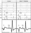
Figure 2. Notch inhibition disrupts AV nodal delay.
The top panels show representative 6-lead EKG and intracardiac electrograms from the high right atrium (HRA) and His bundle electrogram (HBE), demonstrating shorter PR and AH intervals in Mlc2vCre/+DNMAML mice when compared with those in control mice. A, atrial depolarization; P, P wave; V, ventricular depolarization. Scale bar: 50 ms. The bottom panels show 1 representative beat from the His bundle electrogram at higher magnification, with the AH interval demarcated by the bars. The control AH interval was 33 ms, and the Mlc2vCre/+DNMAML AH interval was 22 ms. Control mice are DNMAML littermates of Mlc2vCre/+DNMAML mice.
Figure 3
Figure 3. Activation of Notch signaling results in accessory pathway formation.
(A and B) Scanning electron microscopy demonstrates excess tissue in the right AV junction of 1-week-old Mlc2vCre/+NICD mice (arrow in B) when compared with that of control mice, which have a distinct AV groove (arrow in A). Scale bar: 100 μm (A and B). (C and D) Masson’s trichrome staining of postnatal day 3 hearts reveals an epicardial accessory pathway traversing the AV groove in Notch-activated hearts (arrow, D), connecting atrial and ventricular myocardium, while the AV groove from a similar region in control hearts contains no myocardial tissue (arrow, C). Note the presence of myocardial tissue along the atrial side of the tricuspid valve (asterisk, D). (EH) In situ hybridization reveals that accessory pathways express (F) the atrial marker Mlc2a (red) near the atrium and (H) Mlc2v (red) near the ventricle. Arrowheads in E and G denote the AV groove, and arrowheads in F and H denote an epicardial accessory pathway. Scale bar: 100 μm (CH). Control mice are NICD littermates of Mlc2vCre/+NICD mice.
Figure 4
Figure 4. Developmental analysis reveals late gestation AV canal defects in Mlc2vCre/+NICD embryos.
In situ hybridization of staged embryos from E10.5–E18.5 was performed to determine the earliest signs of detectable AV canal defects in Mlc2vCre/+NICD specimens. (A and B) Bmp2 is expressed in AV canal myocardium of control embryos at E10.5 and E12.5, and (F and G) expression is unchanged in mutants. (C and D) Tbx3 marks AV canal myocardium in the posterior region of the heart at E15.5 and E17.5 and reveals a sharp boundary of expression between the thin rim of remaining AV canal myocardium and ventricular tissue in controls. (H and I) In mutant embryos, Tbx3 expression is slightly expanded and the boundary is irregular, especially at E17.5 (arrows, I). (E and J) MF-20 antibody staining for myosin expression at E16.5 identifies coronary veins of control embryos in the AV groove (asterisk in E). In mutant embryos, the coronary veins were enlarged and were surrounded by a muscular wall expressing myosin (asterisk in J). Arrowheads denote the AV canal in A, B, F, and G, and arrows denote the boundary between AV canal tissue and ventricular tissue in C, D, H, and I. Scale bar: 500 μm (AD and FI); 50 μm (E and J). Control embryos are NICD littermates of Mlc2vCre/+NICD embryos.
Figure 5
Figure 5. Notch activation leads to ventricular preexcitation.
Representative 6-lead EKG tracings and intracardiac electrograms from the high right atrium and His bundle electrogram are shown from a control and Mlc2vCre/+NICD mouse. Note the fused p wave (P) and QRS complexes and widened QRS with delta wave on surface EKG as well as the fused atrial (A) and ventricular (V) electrograms in Notch-activated mice, indicative of robust AV conduction over the accessory pathway. Scale bar: 100 ms. Control mice are NICD littermates of Mlc2vCre/+NICD mice.
Figure 6
Figure 6. Activation of Notch signaling leads to ventricular preexcitation.
Electrical activation maps from the posterior surface of the heart, with 1-ms contour lines for (A and B) control and (C and D) Mlc2vCre/+NICD hearts. Note the different time scales. Atrial activation in control hearts is followed by a significant AV delay prior to ventricular activation. In Notch-activated hearts, the electrical impulse travels from the atria into the ventricles without AV delay via (C) an accessory pathway along the right posterior surface or via (D) dual accessory pathways in the right and left posterior regions, resulting in abnormal activation of the ventricles from base to apex. (E and F) Still frame activation images of the hearts from B and D, taken every 5 ms, in which a time gap of 45 ms, corresponding to AV delay, is denoted in E by a change from yellow to red numbers. White tissue is depolarized. RA, right atrium; LA, left atrium. Control mice are NICD littermates of Mlc2vCre/+NICD mice.

Comment in

References

    1. Bakker ML, Christoffels VM, Moorman AF. The cardiac pacemaker and conduction system develops from embryonic myocardium that retains its primitive phenotype. J Cardiovasc Pharmacol. 2010;56(1):6–15. doi: 10.1097/FJC.0b013e3181e775d3. - DOI - PubMed
    1. Christoffels VM, Smits GJ, Kispert A, Moorman AF. Development of the pacemaker tissues of the heart. Circ Res. 2010;106(2):240–254. doi: 10.1161/CIRCRESAHA.109.205419. - DOI - PubMed
    1. Aanhaanen WT, et al. The Tbx2+ primary myocardium of the atrioventricular canal forms the atrioventricular node and the base of the left ventricle. Circ Res. 2009;104(11):1267–1274. doi: 10.1161/CIRCRESAHA.108.192450. - DOI - PubMed
    1. Horsthuis T, et al. Gene expression profiling of the forming atrioventricular node using a novel tbx3-based node-specific transgenic reporter. Circ Res. 2009;105(1):61–69. doi: 10.1161/CIRCRESAHA.108.192443. - DOI - PubMed
    1. Kokubo H, Tomita-Miyagawa S, Hamada Y, Saga Y. Hesr1 and Hesr2 regulate atrioventricular boundary formation in the developing heart through the repression of Tbx2. Development. 2007;134(4):747–755. doi: 10.1242/dev.02777. - DOI - PubMed

Publication types

MeSH terms

Substances