. 2010:1:147.
doi: 10.1038/ncomms1146.

Cryptic prophages help bacteria cope with adverse environments

Affiliations
Free PMC article

Cryptic prophages help bacteria cope with adverse environments

Xiaoxue Wang et al. Nat Commun. 2010.
Free PMC article

Abstract

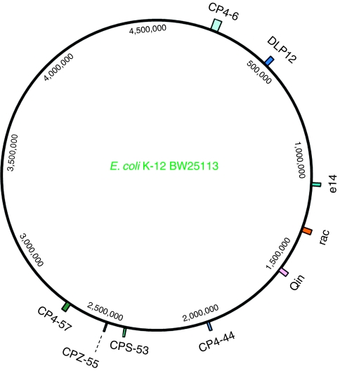
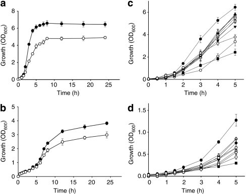
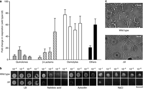
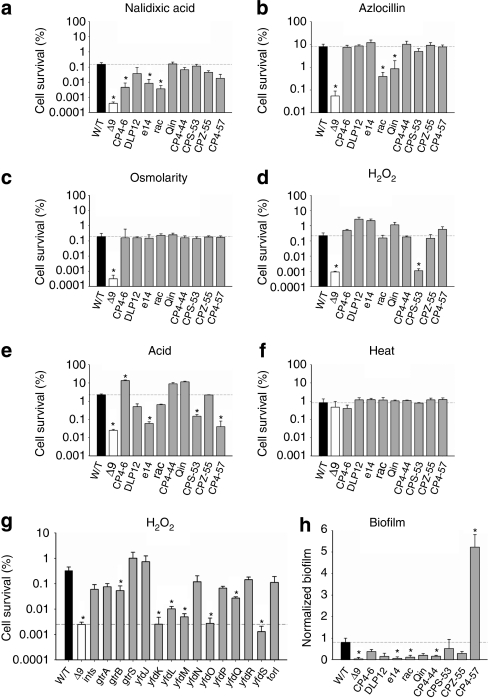
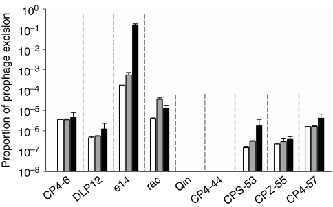
Phages are the most abundant entity in the biosphere and outnumber bacteria by a factor of 10. Phage DNA may also constitute 20% of bacterial genomes; however, its role is ill defined. Here, we explore the impact of cryptic prophages on cell physiology by precisely deleting all nine prophage elements (166 kbp) using Escherichia coli. We find that cryptic prophages contribute significantly to resistance to sub-lethal concentrations of quinolone and β-lactam antibiotics primarily through proteins that inhibit cell division (for example, KilR of rac and DicB of Qin). Moreover, the prophages are beneficial for withstanding osmotic, oxidative and acid stresses, for increasing growth, and for influencing biofilm formation. Prophage CPS-53 proteins YfdK, YfdO and YfdS enhanced resistance to oxidative stress, prophages e14, CPS-53 and CP4-57 increased resistance to acid, and e14 and rac proteins increased early biofilm formation. Therefore, cryptic prophages provide multiple benefits to the host for surviving adverse environmental conditions.


Figures

Figure 1
Figure 1. Size and position of nine cryptic prophages in the E. coli K-12 BW25113 genome.
Map of the E. coli K-12 BW25113 genome demonstrating the cyptic prophages deleted in this study.
Figure 2
Figure 2. Cryptic prophage genes influence cell growth.
Growth in LB at 37 °C (a) and M9C at 30 °C (b) for Δ9 (open circle) and the wild-type strain (filled circle). Growth of the nine individual prophage deletion strains (CP4-6, open diamond; DLP12, filled diamond; e14, open hexagon; rac, inverted open triangle; Qin, filled triangle; CPS-53, inverted filled triangle; CP4-44, open square; CPZ-55, open triangle and CP4-57, filled square), Δ9 (open circle) and the wild-type strain (filled circle) in LB at 37 °C (c) and M9C lactate at 37 °C (d). Error bars indicate s.d. values (n=3).
Figure 3
Figure 3. Cryptic prophage genes influence stress-related phenotypes.
(a) Summary of the PM results for the wild-type strain versus Δ9. The fold changes indicate the relative difference in the average slope in the growth curves multiplied by the area under the curves, which is indicative of cell respiration. From left to right, with the MIC indicated for the wild-type strain in parenthesis, quinolones include nalidixic acid (4 μg ml−1), oxolinic acid (0.9 μg ml−1), ofloxacin (0.048 μg ml−1) and novobiocin (132 μg ml−1); β-lactams include nafcillin (320 μg ml−1), azlocillin (17.6 μg ml−1), cephalothin (5.12 μg ml−1) and moxalactam (0.27 μg ml−1); osmolytes include 5% NaCl, 6% NaCl, 6% KCl and 2% sodium formate; and the other stresses include thallium acetate and potassium tellurite. All chemicals shown here were significantly repressed for Δ9 versus wild-type strain (P<0.05 using a paired t-test). Error bars indicate s.e.m. (n=3). (b) Efficiency of colony formation for Δ9 and the wild-type strain with sublethal concentrations of nalidixic acid (2 μg ml−1) and azlocillin (5 μg ml−1) as well as with 6% NaCl. Powers of 10 indicate the amount of dilution. Scale bar represents 10 mm. (c) Cell morphology for Δ9 and wild-type strain after treating with 2 μg ml−1 nalidixic acid for 2 h. Scale bar represents 10 μm.
Figure 4
Figure 4. Individual prophages affect cell viability with stress and biofilm formation.
Survival for the nine single-prophage deletion strains (marked with each prophage name) and the multiple prophage-deletion strain (Δ9) after challenging with (a) nalidixic acid (2 μg ml−1 for 12 h), (b) azlocillin (4 μg ml−1 for 12 h), (c) osmotic stress (6% NaCl for 12 h), (d) oxidative stress (30 mM H2O2 for 15 min), (e) acid stress (pH 2.5 for 30 min) and (f) heat stress (65 °C for 10 min). (g) Survival for the 15 single-gene deletion strains of CPS-53 (marked with each gene deletion) and the multiple prophage-deletion strain (Δ9) after challenging with oxidative stress (30 mM H2O2 for 15 min). (h) Normalized biofilm formation in 96-well polystyrene plates in M9C medium after 8 h at 30 °C. Error bars indicate s.d. values (n=4). Significant changes are marked with an asterisk for P<0.05.
Figure 5
Figure 5. Prophage excised at different frequencies.
Proportion of wild-type cells that have the indicated prophage excised without mitomycin C (open bar), with the addition of 1 μg ml−1 of mitomycin C (grey bar) and 10 μg ml−1 of mitomycin C (black bar). Mitomycin C was added to exponential-phase cells (turbidity |1.0) in LB medium at 37 °C for 4 h. Error bars indicate s.d. values (n=3).

References

    1. Lwoff A. Interaction among virus, cell, and organism. Science 152, 1216–1220 (1966). - PubMed
    1. Casjens S. Prophages and bacterial genomics: what have we learned so far? Mol. Microbiol. 49, 277–300 (2003). - PubMed
    1. Edlin G., Lin L. & Bitner R. Reproductive fitness of P1, P2, and Mu lysogens of Escherichia coli. J. Virol. 21, 560–564 (1977). - PMC - PubMed
    1. Canchaya C., Proux C., Fournous G., Bruttin A. & Brüssow H. Prophage genomics. Microbiol. Mol. Biol. Rev. 67, 238–276 (2003). - PMC - PubMed
    1. Lawrence J. G. & Ochman H. Molecular archaeology of the Escherichia coli genome. Proc. Natl Acad. Sci. USA 95, 9413–9417 (1998). - PMC - PubMed

Publication types

Associated data