. 2011 Jan 18;9(1):e1000581.
doi: 10.1371/journal.pbio.1000581.

SecM-stalled ribosomes adopt an altered geometry at the peptidyl transferase center

Affiliations

SecM-stalled ribosomes adopt an altered geometry at the peptidyl transferase center

Shashi Bhushan et al. PLoS Biol. .

Abstract

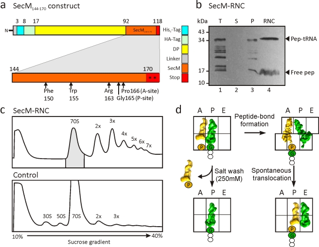
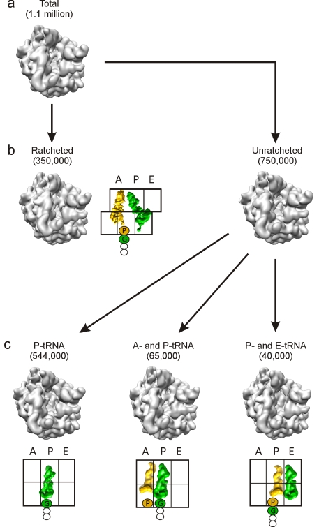
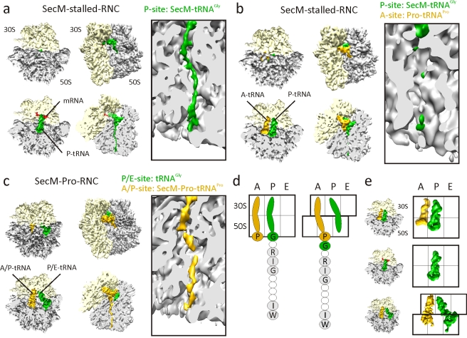
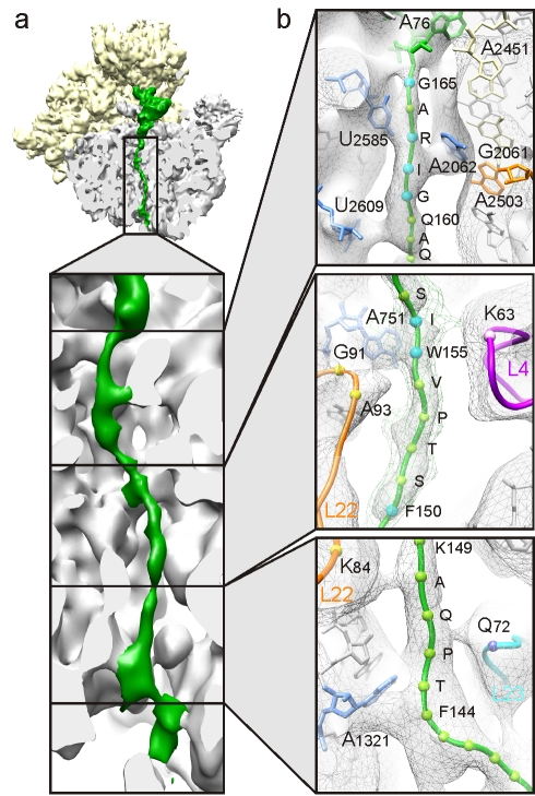
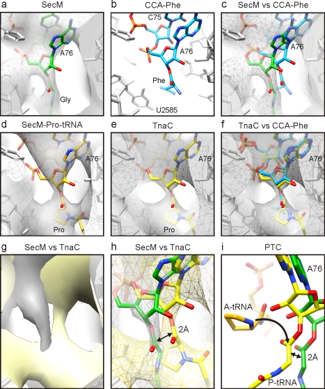
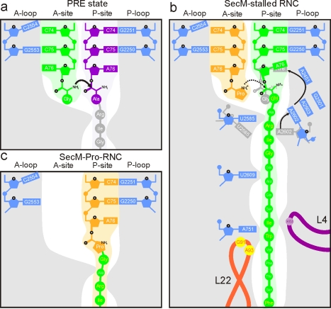
As nascent polypeptide chains are synthesized, they pass through a tunnel in the large ribosomal subunit. Interaction between specific nascent chains and the ribosomal tunnel is used to induce translational stalling for the regulation of gene expression. One well-characterized example is the Escherichia coli SecM (secretion monitor) gene product, which induces stalling to up-regulate translation initiation of the downstream secA gene, which is needed for protein export. Although many of the key components of SecM and the ribosomal tunnel have been identified, understanding of the mechanism by which the peptidyl transferase center of the ribosome is inactivated has been lacking. Here we present a cryo-electron microscopy reconstruction of a SecM-stalled ribosome nascent chain complex at 5.6 Å. While no cascade of rRNA conformational changes is evident, this structure reveals the direct interaction between critical residues of SecM and the ribosomal tunnel. Moreover, a shift in the position of the tRNA-nascent peptide linkage of the SecM-tRNA provides a rationale for peptidyl transferase center silencing, conditional on the simultaneous presence of a Pro-tRNA(Pro) in the ribosomal A-site. These results suggest a distinct allosteric mechanism of regulating translational elongation by the SecM stalling peptide.


Conflict of interest statement

The authors have declared that no competing interests exist.

Figures

Figure 1
Figure 1. Generation of SecM-stalled RNCs.
(A) Schematic showing the construct used for translation. The SecM stalling window (residues 144–170, orange) was inserted after the DP sequence and flanked by tandem stop codons (asterisks). Critical residues for stalling are marked by arrows. (B) Western blot (using anti-HA antibody) of SDS-PAGE of the translation reaction (T, lane 1), supernatant (S, lane 2), and pellet (P, lane 3) fractions following centrifugation, as well as the purified RNC following affinity column (RNC, lane 4). The position of the peptidyl-tRNA (pep-tRNA) and free SecM peptide (free pep) are indicated. (C) Sucrose-gradient profiles of the SecM RNCs (after Co-NTA purification) (upper panel) and control translation extract without template (lower panel). The shaded 70S monosomes were collected and pelleted for cryo-EM reconstruction. (D) SecM stalling occurs with SecM-tRNAGly located at the P-site of the ribosome and Pro-tRNAPro at the A-site ,. During purification of the SecM RNC, the Pro-tRNAPro in the A-site can dissociate because of high salt washing, or undergo slow peptide bond formation and form a ratcheted hybrid state –, with SecM-Pro-tRNAPro in the A/P-site and deacylated tRNAGly in the P/E-site. The hybrid state may spontaneously translocate, albeit slowly ,, to form an unratcheted post-state with SecM-Pro-tRNAPro in the P-site and deacylated tRNAGly in the E-site.
Figure 2
Figure 2. Schematic for in silico sorting of the SecM RNCs.
The unsorted volume (A) containing a total of 1.1 million particles with density in all three tRNA binding sites was initially sorted into two populations (B) based on the ratchet-like subunit rearrangement of the small subunit relative to the large subunit. The ratcheted population (350,000 particles; 32%) had tRNAs present in A/P- and P/E-sites, whereas the unratcheted population (750,000 particles; 68%) could be further sorted into three subpopulations (C): a dominant fraction (544,000 particles; 73%) with P-tRNA only, and two minor fractions with A- and P-tRNAs (65,000; 12%) and with P- and E-tRNAs (40,000; 7%).
Figure 3
Figure 3. Cryo-EM reconstructions of SecM RNCs.
(A–C) Cryo-EM reconstructions of (A) SecM-stalled RNC with SecM-tRNAGly (green) in P-site, (B) SecM-stalled RNC with additional Pro-tRNAPro (gold) in the A-site, and (C) SecM-Pro-RNC, with SecM-Pro-tRNAPro (gold) in A/P-site and tRNAGly (green) in P/E-site. For each reconstruction, the top two diagrams show a top and factor view of the small (30S, yellow) and large (50S, gray) subunits, with respective cross-sections below. Right-hand panels show close-up of the tunnel views of each complex. (D) Schematic showing unratcheted SecM-stalled state (left), with Pro-tRNAPro in the A-site and SecM-tRNAGly in the P-site, and post-arrest ratcheted state (right), with SecM-Pro-tRNAPro in the hybrid A/P-site and tRNAGly in the P/E-site. Residues important for SecM stalling are shaded and labeled with single-letter amino acid code. (E) Schematic showing the relative positions of the tRNAs from the complexes in (A–C).
Figure 4
Figure 4. SecM nascent chain interactions with tunnel components.
(A) Cross-section of the large ribosomal subunit of the SecM-stalled RNC revealing the sites of interaction between the SecM nascent chain (green) and the ribosomal tunnel (gray). (B) Close-up of the upper, middle, and lower regions of the ribosomal tunnel with density (gray mesh) and molecular models for SecM nascent chain (green, with balls marking the Ca of the labeled residues; blue indicates the residue is important for stalling), the 23S rRNA (gray, except for selected colored nucleotides), and ribosomal proteins L4 (purple), L22 (orange), and L23 (cyan).
Figure 5
Figure 5. Inactivation of the ribosomal PTC by SecM stalling.
(A–C) Identical views of the position of (A) the SecM-tRNAGly (green) in the map (gray mesh) of the SecM-stalled RNC and (B) CCA-Phe (cyan) based on the crystal structure of the archaeal large ribosomal subunit in complex with CCA-PCB (PDB ID1VQN) . (C) Comparison of (A) and (C). (D–F) Identical views of the position of (D) Pro-tRNAPro (orange) in the map (gray mesh) of the SecM-Pro-RNC and (E) TnaC-tRNAPro (yellow) in the map (gray mesh) of the TnaC-stalled RNC. (F) Comparison of (B) and (E). (G and H) Comparison of the SecM- (gray) and TnaC-stalled RNC (yellow) maps as surfaces (G) and as mesh with molecular models for SecM-tRNAGly (green) and TnaC-tRNAPro (yellow) (H). (I) Position of Pro-tRNAPro (A-tRNA, orange; derived from PDB ID1VQN) relative to TnaC-tRNAPro (yellow) and SecM-tRNAGly (green). The arrow indicates the nucleophilic attack of the α-amino of the A-tRNA on the carbonyl carbon of the P-tRNA, which is displaced by 2 Å in the SecM-stalled RNC.
Figure 6
Figure 6. Schematic for SecM action.
(A) Schematic representation showing canonical peptide bond formation where the CCA-ends of the tRNAs are precisely positioned to promote nucleophilic attack of the carbonyl carbon of the P-tRNA (purple) by the α-amino of the A-tRNA (green). (B) Interaction of the SecM nascent chain with components of the tunnel aids in the positioning of the critical Arg163, which interacts with A2062 of the 23S rRNA. Interaction of A2062 with A2503 has been proposed to trigger a relay that leads to inactivation of the PTC . We propose that this results from a shifted position of the A76 of the SecM-tRNAGly in the P-site, which prevents efficient attack of the A-tRNA. (C) During prolonged SecM stalling, or by SecA activity, release from the arrested state occurs. The SecM-Pro-tRNAPro forms through peptide bond formation and can now adopt an A/P hybrid state.

References

    1. Polacek N, Mankin A. S. The ribosomal peptidyl transferase center: structure, function, evolution, inhibition. Crit Rev Biochem Mol Biol. 2005;40:285–311. - PubMed
    1. Simonovic M, Steitz T. A. A structural view on the mechanism of the ribosome-catalyzed peptide bond formation. Biochim Biophys Acta. 2009;1789:612–623. - PMC - PubMed
    1. Becker T, Bhushan S, Jarasch A, Armache J. P, Funes S, et al. Structure of monomeric yeast and mammalian Sec61 complexes interacting with the translating ribosome. Science. 2009;326:1369–1373. - PMC - PubMed
    1. Seidelt B, Innis C. A, Wilson D. N, Gartmann M, Armache J. P, et al. Structural insight into nascent polypeptide chain-mediated translational stalling. Science. 2009;326:1412–1415. - PMC - PubMed
    1. Bhushan S, Gartmann M, Halic M, Armache J. P, Jarasch A, et al. Alpha-helical nascent polypeptide chains visualized within distinct regions of the ribosomal exit tunnel. Nat Struct Mol Biol. 2010;17:313–317. - PubMed

Publication types

MeSH terms