. 2010 Nov 17:4:208.
doi: 10.3389/fnhum.2010.00208. eCollection 2010.

Economic value biases uncertain perceptual choices in the parietal and prefrontal cortices

Affiliations

Economic value biases uncertain perceptual choices in the parietal and prefrontal cortices

Christopher Summerfield et al. Front Hum Neurosci. .

Abstract

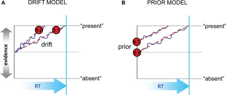
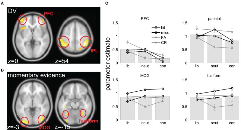
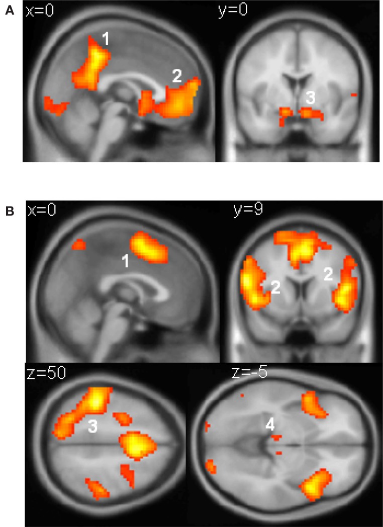
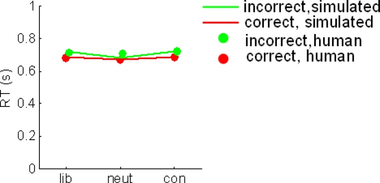
An observer detecting a noisy sensory signal is biased by the costs and benefits associated with its presence or absence. When these costs and benefits are asymmetric, sensory, and economic information must be integrated to inform the final choice. However, it remains unknown how this information is combined at the neural or computational levels. To address this question, we asked healthy human observers to judge the presence or absence of a noisy sensory signal under economic conditions that favored yes responses (liberal blocks), no responses (conservative blocks), or neither response (neutral blocks). Economic information biased fast choices more than slow choices, suggesting that value and sensory information are integrated early in the decision epoch. More formal simulation analyses using an Ornstein-Uhlenbeck process demonstrated that the influence of economic information was best captured by shifting the origin of evidence accumulation toward the more valuable bound. We then used the computational model to generate trial-by-trial estimates of decision-related evidence that were based on combined sensory and economic information (the decision variable or DV), and regressed these against fMRI activity recorded whilst participants performed the task. Extrastriate visual regions responded to the level of sensory input (momentary evidence), but fMRI signals in the parietal and prefrontal cortices responded to the decision variable. These findings support recent single-neuron data suggesting that economic information biases decision-related signals in higher cortical regions.

Keywords: bias; computational modeling; fMRI; parietal cortex; perceptual decision-making; prefrontal cortex; reward.


Figures

Figure 1
Figure 1
Schematic representation of the drift and prior models. (A) In the drift model, decisions are biased toward the more valuable option (here, “present”) by increasing the rate of evidence accumulation. Values inside the red circles show representative gain values; blue trace and red dashed trace show evidence accumulation with and without noise. Evidence toward “present” or “absent” is represented on the vertical axis, and time (RT or cycles) on the horizontal axis. The bound is represented with dashed gray lines top and bottom; another dashed line signals the equilibrium point between choices. RT for the slower of the two scenarios is shown with a cyan vertical line. (B) In the prior model, the initial estimates of evidence in favor of the more valuable option are increased, even before the onset of accumulation. Values in the red circles denote possible prior values in units of probability (i.e., p = 0.5 reflects equilibrium between the two choices). Otherwise as (A).
Figure 2
Figure 2
Task and behavioral data. (A) Experimental paradigm. Each block consisted of 8 (or 16 in behavioral sessions) successive presentations of low-contrast Gabor patches in visual noise. A colored frame and tint to the stimulus denoted bias condition (here, blue). Subjects received auditory feedback at the offset of each trial, and each block closed with a bonus screen informing the subject of their winnings/losses. In behavioral sessions on days 1 and 2, subjects additionally received visual feedback in the form of an advancing or receding “payment bar” immediately underneath the stimulus. (B) Payoff matrices for each condition. Subjects received the indicated values for hits, misses, false alarms (FA) and correct rejections (CR). (C) Estimates of criterion (c) for each individual subject in liberal (red dots), neutral (green dots), and conservative (blue dots) blocks. Black dashed line represents c = 0, i.e., no bias. (D) False alarm rate plotted as a function of reaction time quantile for liberal (red lines), neutral (green lines) and conservative (blue lines) blocks. Bars are standard error of the mean (SEM). (E). Criterion (c) plotted as a function of reaction time quantile for liberal (red lines), neutral (green lines), and conservative (blue lines) blocks. Bars are standard error of the mean (SEM).
Figure 3
Figure 3
(A) Fits of the prior model to human hit rates (left panel) and false alarm (FA) rates (right panel), plotted for RT quartiles (fastest 25, 25–50, 50–75, and >75%) in liberal (red), neutral (green), and conservative (blue) conditions. Circles plot the mean of human subject data; lines plot the simulated data from each model variant. (B) Similarly, fits for the drift model (bottom panel) to hit rates and FA rates. Dashed red boxes highlight the fast, liberal false alarms, which are poorly fit by this model.
Figure 4
Figure 4
(A) Human subject (top panels) and simulated (bottom panels) RT distributions for hits (orange lines), misses (pink lines), false alarms (cyan lines), and correct rejections (brown lines) in liberal (left panels), neutral (middle panels), and conservative (right panels) conditions. RT distributions are normalized to reflect proportions of trials. (B) Example simulated evidence accumulation traces from the best-fitting model (signal-present trials only) in liberal (left panel), neutral (middle panel), and conservative (right panel) conditions. Each trace represents one simulated trial; the thicker trace is the median of all trials. Simulated time is shown on the x-axis and evidence (I) on the y-axis. Dashed lines indicate upper and lower bounds (for “yes” and “no” respectively) and the central dashed line represents equilibrium between the two choices. Traces bend toward either axis because the best-fitting model included an attractor value (λ) of 0.05. (C) Scatter plot of simulated α (y-axis) with RT under the parameters of the best-fitting model, for hits (top left panel), misses (top right panel), FA (bottom left), and CR (bottom right) trials. The black line shows the best fit of a 3rd order polynomial to the data for each trial type.
Figure 5
Figure 5
Brain imaging results. (A) axial views of voxels responding to the DV rendered onto a standard brain in the space of the Montreal Neurological Institute (MNI), at an FDR-corrected threshold of p < 0.05. Slices are labeled with their coordinates in the z plane at the bottom left-hand corner. (B) A comparable plot for voxels responding to momentary evidence. (C) Parameter estimates (from factorial ANOVA analyses) for hits, misses, FA and CR trials in liberal, neutral and conservative blocks, averaged across voxels in each of the four clusters (prefrontal cortex – PFC; intraparietal lobule – IPL; middle occipital gyrus – MOG, and fusiform gyrus. Gray lines are for signal absent trials, black lines for signal present trials; full lines for “yes” responses, dashed lines for “no” responses. Underlying bars illustrate mean parameter estimates for liberal, neutral and conservative blocks.
Figure S1
Figure S1
(A) Voxels responding to reward. Responses that scaled with reward obtained (−5, −3, −1, 1, 3, or 5 points) were observed in (1) the posterior cingulate cortex (peak: −3, −45, 48, T(20) = 12.84), (2) the ventromedial prefrontal cortex (peak: 0, 45, −9, T = 7.39), and (3) the ventral striatum (peak right: 12, 6, −15, T(20) = 8.26; peak left: −12, 6, −15, T(20) = 7.13), as well as in the visual cuneus and lateral OFC. (B) Voxels responding to parametrically with increasing reaction time. These were found in (1) the SMA/preSMA (peak: −3, 6, 51, T(20) = 9.09), (2) the anterior insular cortex (peak right: 36, 21, 3, T(20) = 10.1; peak left: −48, 12, 0, T(20) = 9.45), (3) the parietal (peak: −45, −33, 54, T(20) = 12.98) and premotor (peak: −24, −6, 60; T(20) = 10.72) cortices predominantly on the left, and the (4) the thalamus (peak left: −9, −18, 6, T(20) = 5.84; peak right: 9, −18, 9, T(20) = 7.2) as well as in more dorsal prefrontal regions. All voxels reported in this figure survive an uncorrected threshold of at least p < 1 × 10−5).
Figure S2
Figure S2
Correct (red dots) and incorrect (green dots) reaction times in liberal, neutral and conservative conditions. Lines show the best fits to these data from the prior model.

References

    1. Anderson A. J., Yadav H., Carpenter R. H. (2008). Directional prediction by the saccadic system. Curr. Biol. 18, 614–61810.1016/j.cub.2008.03.057 - DOI - PubMed
    1. Bar M., Kassam K. S., Ghuman A. S., Boshyan J., Schmid A. M., Dale A. M., Hamalainen M. S., Marinkovic K., Schacter D. L., Rosen B. R., Halgren E. (2006). Top-down facilitation of visual recognition. Proc. Natl. Acad. Sci. U.S.A. 103, 449–45410.1073/pnas.0507062103 - DOI - PMC - PubMed
    1. Bar M., Tootell R. B., Schacter D. L., Greve D. N., Fischl B., Mendola J. D., Rosen B. R., Dale A. M. (2001). Cortical mechanisms specific to explicit visual object recognition. Neuron 29, 529–53510.1016/S0896-6273(01)00224-0 - DOI - PubMed
    1. Bogacz R., Brown E., Moehlis J., Holmes P., Cohen J. D. (2006). The physics of optimal decision making: a formal analysis of models of performance in two-alternative forced-choice tasks. Psychol. Rev. 113, 700–76510.1037/0033-295X.113.4.700 - DOI - PubMed
    1. Boorman E. D., Behrens T. E., Woolrich M. W., Rushworth M. F. (2009). How green is the grass on the other side? Frontopolar cortex and the evidence in favor of alternative courses of action. Neuron 62, 733–74310.1016/j.neuron.2009.05.014 - DOI - PubMed

LinkOut - more resources