Review
. 2011 Jun;36(3):264-72.
doi: 10.1007/s00261-011-9685-1.

Hepatocelluar nodules in liver cirrhosis: hemodynamic evaluation (angiography-assisted CT) with special reference to multi-step hepatocarcinogenesis

Affiliations
Review

Hepatocelluar nodules in liver cirrhosis: hemodynamic evaluation (angiography-assisted CT) with special reference to multi-step hepatocarcinogenesis

Osamu Matsui et al. Abdom Imaging. 2011 Jun.

Abstract

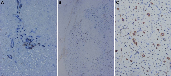
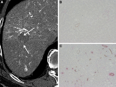
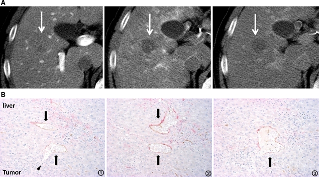
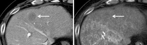
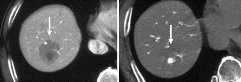
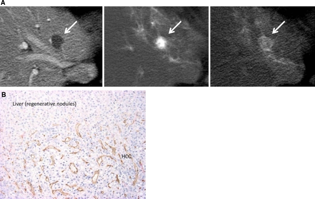
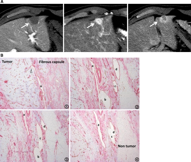
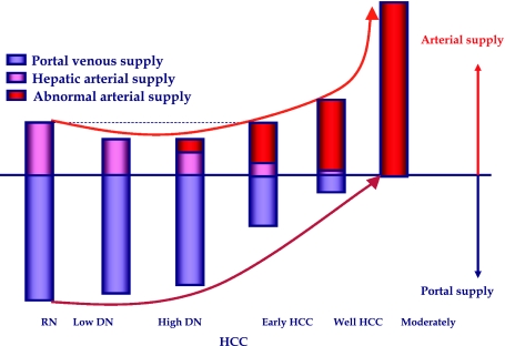
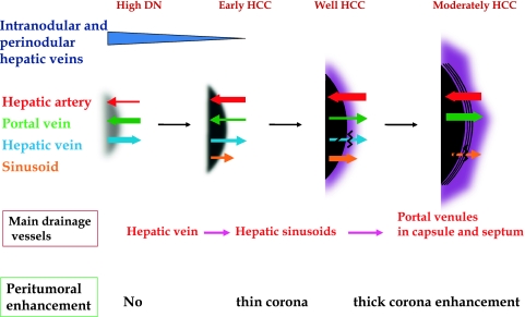
To understand the hemodynamics of hepatocellular carcinoma (HCC) is important for the precise imaging diagnosis and treatment, because there is an intense correlation between their hemodynamics and pathophysiology. Angiogenesis such as sinusoidal capillarization and unpaired arteries shows gradual increase during multi-step hepatocarcinogenesis from high-grade dysplastic nodule to classic hypervascular HCC. In accordance with this angiogenesis, the intranodular portal supply is decreased, whereas the intranodular arterial supply is first decreased during the early stage of hepatocarcinogenesis and then increased in parallel with increasing grade of malignancy of the nodules. On the other hand, the main drainage vessels of hepatocellular nodules change from hepatic veins to hepatic sinusoids and then to portal veins during multi-step hepatocarcinogenesis, mainly due to disappearance of the hepatic veins from the nodules. Therefore, in early HCC, no perinodular corona enhancement is seen on portal to equilibrium phase CT, but it is definite in hypervascular classical HCC. Corona enhancement is thicker in encapsulated HCC and thin in HCC without pseudocapsule. To understand these hemodynamic changes during multi-step hepatocarcinogenesis is important, especially for early diagnosis and treatment of HCCs.


Figures

Fig. 1
Fig. 1
Angiogenesis during multi-step hepatocarcinogenesis. Sinusoidal capillarization and unpaired arteries are expressed from spotty in DN (A, ×100), diffuse in early hepatocellular carcinoma (HCC) (B, ×50) to intense distribution in moderately differentiated HCC (C, ×100) in accordance with the elevation of the grade of malignancy of the nodules during hepatocarcinogenesis [Double immunohistochemical staining for CD 34 (blue) and α-smooth muscle actin (SMA) (brown)].
Fig. 2
Fig. 2
Early HCC (high-grade DN with a well-differentiated HCC focus). A CT during hepatic arteriography (CTHA) shows entirely hypodense nodule (arrow b) with a slightly hyperdense focus (arrow c). B Double immunohistochemical staining for CD34 and αSMA of DN portion (arrow b in A) shows no definite expression of sinusoidal capillarization and unpaired arteries. C Double immunohistochemical staining of HCC portion corresponding (arrow c in A) shows expression of sinusoidal capillarization and unpaired arteries.
Fig. 3
Fig. 3
High-grade DN. A Early phase of CTHA shows a hypodense nodule (middle arrow). It demonstrates isodensity on CT during arterial portography (CTAP, left arrow) and no perilesional enhancement on late phase of CTHA (right arrow). B Serial specimens from 1 to 3 with double immunohistochemical staining for CD 34 and α-SMA show the communication between intratumoral blood sinusoids (arrowhead) and hepatic venules (arrows) in the tumor (×100).
Fig. 4
Fig. 4
Early HCC. CTAP (left) shows a faintly hypodense nodule indicating decreased but not absent intranodular portal supply (arrow). On CTHA (right), it is not well identified indicating almost the same intranodular arterial supply relative to the surrounding liver (arrow).
Fig. 5
Fig. 5
Well-differentiated HCC (early HCC with subfocus of definite hypervascular well-differentiated HCC). CTAP (left) shows a slightly hypodense nodule with an internal definitely hypodense focus indicating partial portal perfusion defect (arrow). On CTHA (right), it is visualized as an entirely isodense nodule with an internal definitely hypervascular focus (arrow).
Fig. 6
Fig. 6
Hypervascular well-differentiated HCC. A CTAP (left) shows an entirely hypodense nodule (arrow). It demonstrates hyperdensity on early phase of CTHA (middle, arrow) and thin corona enhancement on late phase of CTHA (right, arrow). B Double immunohistochemical staining of the boundary between the tumor and liver parenchyma shows abundant communications between intratumoral blood sinusoids and hepatic sinusoids (×100).
Fig. 7
Fig. 7
Moderately differentiated HCC with pseudocapsule formation. A CTAP shows an entirely hypodense nodule (left, arrow). CTHA demonstrates it as an entirely hyperdense nodule on early phase (middle, arrow) with thick corona enhancement on late phase (right, arrow). B Serial specimens from 1 to 4 with double immunohistochemical staining for CD34 and αSMA show the communication between intra-capsular portal venules (ad) and intratumoral blood sinusoids (arrowheads) (×100).
Fig. 8
Fig. 8
Multi-step hepatocarcinogenesis and changes of intranodular blood supply. Intranodular portal supply gradually decreases in accordance with the elevation of the grade of malignancy of the nodules and finally disappears in moderately differentiated HCCs. On the other hand, arterial supply first decreases at the early stage of hepatocarcinogenesis and then acutely increases, and finally the entire nodule is fed only by artery in moderately differentiated HCCs.
Fig. 9
Fig. 9
Multi-step changes of drainage vessels and peritumoral enhancement during hepatocarcinogenesis. In DNs or early HCCs, the main drainage route from the tumor is intranodular or perinodular hepatic vein. However, because hepatic veins disappear from the tumor during very early stage of hepatocarcinogenesis, drainage vessels change to hepatic sinusoids. In moderately differentiated HCC with pseudocapsule formation, the communication between tumor sinusoids and the surrounding hepatic sinusoids are also blocked, and then, the portal venules in the pseudocapsule finally become the main drainage vessel from the tumor. In accordance with the changes of the drainage vessels, thin to thick corona enhancement appears surrounding the tumor.

References

    1. Ikai I, Arii S, Okazaki M, et al. Report of the 17th nationwide follow-up survey of primary liver cancer in Japan. Hepatol Res. 2007;37:676–691. doi: 10.1111/j.1872-034X.2007.00119.x. - DOI - PubMed
    1. Matsui O, Kadoya M, Suzuki M, et al. Work in progress: dynamic sequential computed tomography during arterial portography in the detection of hepatic neoplasms. Radiology. 1983;146:721–727. - PubMed
    1. Matsui O, Takashima T, Kadoya M, et al. Dynamic computed tomography during arterial portography: the most sensitive examination for small hepatocellular carcinomas. J Comput Assist Tomogr. 1985;9:19–24. doi: 10.1097/00004728-198501000-00004. - DOI - PubMed
    1. Hayashi M, Matsui O, Ueda K, et al. Correlation between the blood supply and grade of malignancy of hepatocellular nodules associated with liver cirrhosis: evaluation by CT during intraarterial injection of contrast medium. AJR Am J Roentgenol. 1999;172:969–976. - PubMed
    1. International Consensus Group for Hepatocellular Neoplasia Pathologic diagnosis of early hepatocellular carcinoma: a report of the international consensus group for hepatocellular neoplasia. Hepatology. 2009;49:658–664. doi: 10.1002/hep.22709. - DOI - PubMed

MeSH terms