. 2011 Jan 26:12:26.
doi: 10.1186/1471-2474-12-26.

Second harmonic generation analysis of early Achilles tendinosis in response to in vivo mechanical loading

Affiliations

Second harmonic generation analysis of early Achilles tendinosis in response to in vivo mechanical loading

Thomas Abraham et al. BMC Musculoskelet Disord. .

Abstract

Background: Tenocytes have been implicated in the development of tendinosis, a chronic condition commonly seen in musculoskeletal overuse syndromes. However, the relation between abnormal tenocyte morphology and early changes in the fibrillar collagen matrix has not been closely examined in vivo. Second harmonic generation (SHG) microscopy is a recently developed technique which allows examination of fibrillar collagen structures with a high degree of specificity and resolution. The goal of this study was to examine the potential utility of SHG and multiphoton excitation fluorescence (MPEF) microscopy in understanding the relation between tenocytes and their surrounding collagenous matrix in early tendon overuse lesions.

Methods: Histological preparations of tendon were prepared from adult male Sprague-Dawley rats subjected to an Achilles tendon loading protocol for 12 weeks (Rat-A-PED), or from sedentary age-matched cage controls. Second harmonic generation and multiphoton excitation fluorescence were performed simultaneously on these tissue sections in at least three different areas.

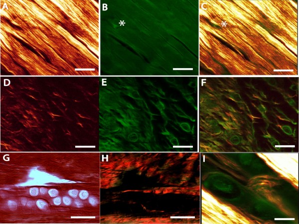
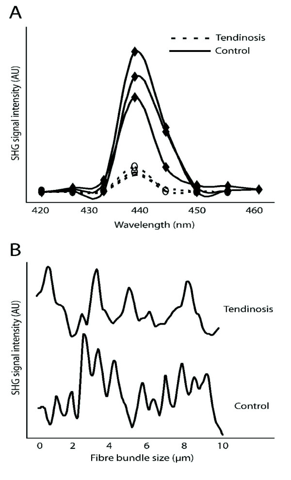
Results: SHG microscopy revealed an association between abnormal tenocyte morphology and morphological changes in the fibrillar collagen matrix of mechanically loaded Achilles tendons. Collagen density and organization was significantly reduced in focal micro-regions of mechanically loaded tendons. These pathological changes occurred specifically in association with altered tenocyte morphology. Normal tendons displayed a regular distribution of fibre bundles, and the average size of these bundles as determined by Gaussian analysis was 0.47 μm ± 0.02. In comparison, fibre bundle measures from tendon regions in the vicinity of abnormal tenocytes could not be quantified due to a reduction in their regularity of distribution and orientation.

Conclusions: SHG microscopy allowed high resolution detection of focal tendon abnormalities affecting the fibrillar collagen matrix. With ongoing repetitive loading, these tenocyte-associated focal collagen defects could predispose to the progression of overuse pathology.


Figures

Figure 1
Figure 1
Multiphoton and SHG signals originating from tendon histological preparations. MPEF images show general tissue morphology of tendon and associated structures, while spectrally clean SHG images specifically reveal collagen present in the same region. Representative SHG (A,D), MPEF (B,E) or combined images (C,F,I) obtained from standard histological thin tissue section are shown. In normal healthy tendon (A-C), SHG demonstrates tightly bundled, longitudinally oriented collagen fibres. Tenocytes (B, C, asterisk) are inconspicuous due to their sparse cytoplasm. In early tendinosis tendon, collagen surrounding abnormal tenocytes (D-F) demonstrates a disturbed organization, and the average SHG signal is greatly reduced (c.f. panel A). The tenocytes in tendinosis tendon demonstrate much more prominent cytoplasm and a rounded, as opposed to spindle-shaped, morphology (E). Picrosirius red-stained tendon from tendinosis tendon (G, brightfield; H, polarized light) is shown as a comparison. Tendinosis regions demonstrate a loss of birefringence (H). In comparison, SHG signal reveals the presence of abnormal pericellular fibrillar collagen structures not visualized with polarized light. Scale bars represent 25 μm for A-H, and 10 μm for I.
Figure 2
Figure 2
Analysis of SHG signal intensity in rat Achilles tendon from collagen surrounding normal or abnormal tenocytes. (A) Three representative spectral scans from a single normal (solid line) and a single tendinosis tendon (dashed line) are shown. The SHG signal arising from the tendon proper region peaked consistently at 440 nm as expected. (B) When SHG signal intensity was plotted as a function of position along a line drawn perpendicular to the collagen fibre bundles, the fibre bundle size could be estimated with sub-micron resolution in normal tendon, but not in tendinosis tendons.

Similar articles

Cited by

References

    1. Abraham T, Carthy J, McManus B. Collagen matrix remodeling in 3-dimensional cellular space resolved using second harmonic generation and multiphoton excitation fluorescence. J Struct Biol. 2010;169:36–44. doi: 10.1016/j.jsb.2009.07.023. - DOI - PubMed
    1. Strupler M, Pena AM, Hernest M, Tharaux PL, Martin JL, Beaurepaire E, Schanne-Klein MC. Second harmonic imaging and scoring of collagen in fibrotic tissues. Opt Express. 2007;15:4054–4065. doi: 10.1364/OE.15.004054. - DOI - PubMed
    1. Zipfel WR, Williams RM, Christie R, Nikitin AY, Hyman BT, Webb WW. Live tissue intrinsic emission microscopy using multiphoton-excited native fluorescence and second harmonic generation. Proc Natl Acad Sci USA. 2003;100:7075–7080. doi: 10.1073/pnas.0832308100. - DOI - PMC - PubMed
    1. Coleman BD, Khan KM, Maffulli N, Cook JL, Wark JD. Studies of surgical outcome after patellar tendinopathy: clinical significance of methodological deficiencies and guidelines for future studies. Victorian Institute of Sport Tendon Study Group. Scand J Med Sci Sports. 2000;10:2–11. doi: 10.1034/j.1600-0838.2000.010001002.x. - DOI - PubMed
    1. Jarvinen M, Jozsa L, Kannus P, Jarvinen TL, Kvist M, Leadbetter W. Histopathological findings in chronic tendon disorders. Scand J Med Sci Sports. 1997;7:86–95. doi: 10.1111/j.1600-0838.1997.tb00124.x. - DOI - PubMed

Publication types

Grants and funding