. 2011 Mar;138(5):915-24.
doi: 10.1242/dev.057729. Epub 2011 Jan 26.

Capicua DNA-binding sites are general response elements for RTK signaling in Drosophila

Affiliations

Capicua DNA-binding sites are general response elements for RTK signaling in Drosophila

Leiore Ajuria et al. Development. 2011 Mar.

Abstract

RTK/Ras/MAPK signaling pathways play key functions in metazoan development, but how they control expression of downstream genes is not well understood. In Drosophila, it is generally assumed that most transcriptional responses to RTK signal activation depend on binding of Ets-family proteins to specific cis-acting sites in target enhancers. Here, we show that several Drosophila RTK pathways control expression of downstream genes through common octameric elements that are binding sites for the HMG-box factor Capicua, a transcriptional repressor that is downregulated by RTK signaling in different contexts. We show that Torso RTK-dependent regulation of terminal gap gene expression in the early embryo critically depends on Capicua octameric sites, and that binding of Capicua to these sites is essential for recruitment of the Groucho co-repressor to the huckebein enhancer in vivo. We then show that subsequent activation of the EGFR RTK pathway in the neuroectodermal region of the embryo controls dorsal-ventral gene expression by downregulating the Capicua protein, and that this control also depends on Capicua octameric motifs. Thus, a similar mechanism of RTK regulation operates during subdivision of the anterior-posterior and dorsal-ventral embryonic axes. We also find that identical DNA octamers mediate Capicua-dependent regulation of another EGFR target in the developing wing. Remarkably, a simple combination of activator-binding sites and Capicua motifs is sufficient to establish complex patterns of gene expression in response to both Torso and EGFR activation in different tissues. We conclude that Capicua octamers are general response elements for RTK signaling in Drosophila.


Figures

Fig. 1.
Fig. 1.
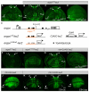
Torso signaling regulates hkb expression via TGAATGAA repressor elements. (A) The hkb locus depicting the hkb0.4 enhancer (red line). EI, EcoRI restriction site located 2.1 kb upstream of the transcription start site. The structure of lacZ reporters is shown below. (B-G) mRNA expression patterns of hkb0.4-lacZ (B-D,F,G) and hkb0.4mut-lacZ (E) in otherwise wild-type (B,E), cic1 (C), cicΔC2 (D), groMB36 (F) and dl1/dl4 (G) embryos. Closed arrowheads in C and F indicate derepressed hkb0.4-lacZ expression in cic1 and groMB36 embryos. Open arrowheads in D and G indicate reduced hkb0.4-lacZ expression in cicΔC2 and dl1/dl4 embryos.
Fig. 2.
Fig. 2.
Cic-binding motifs confer Torso-dependent regulation to synthetic enhancers. (A) Diagram of lacZ reporters containing Bcd-activating sequences and T(G/C)AATGAA sites. The 270 bp hb enhancer (delimited by NheI and MluI restriction sites) is indicated in green. (B-J) mRNA expression patterns of hb-lacZ (B), hbC-lacZ (C,D), Bcd-lacZ (E), CBcdC-lacZ (F-H), CBcdCTRE-lacZ (I) and CBcdCmut-lacZ (J) in otherwise wild-type (B,C,E,F,I,J), cic1 (D,G) and cicΔC2 (H) embryos. Closed arrowheads in D and G indicate expanded hbC-lacZ and CBcdC-lacZ expression in cic1 embryos. The open arrowhead in H indicates residual CBcdC-lacZ expression at the anterior pole.
Fig. 3.
Fig. 3.
Association of Gro with the hkb0.4 enhancer requires intact Cic regulatory sites. (A) The hkb0.4-lacZ transgene contains the 0.4 kb hkb enhancer, which includes two Cic-binding sites, a Dorsal-binding site and a Retn-binding site upstream of the lacZ reporter. The positions of amplicons A-I are shown relative to hkb0.4-lacZ. (B) Crosslinked chromatin was isolated from embryos carrying the hkb0.4-lacZ (blue bars) or the hkb0.4mut-lacZ (red bars) transgenes. Anti-Gro ChIP was assayed by qPCR using amplicons A-I. Each bar represents the average (±s.d.) of three to five independent biological replicates. Background levels resulting from pre-immune ChIP controls were subtracted out of all signals.
Fig. 4.
Fig. 4.
EGFR induces ind expression by relieving Cic repression. (A) The ind locus showing the neighboring Rpn12R gene (predicted to encode a component of the proteasome) and the ind0.5 enhancer present in the 3′-flanking region (blue line). EI, EcoRI site present 4.6 kb downstream of the ind transcription start site. d, Dorsal-binding site (GGGAAATTCCC). lacZ reporters driven by ind0.5 enhancer sequences are also shown. (B-B″) Stage 5 cic-HA; cic1 embryo stained with anti-dpERK (red, B) and anti-HA (green, B′) antibodies; the merged image is shown in B″. EGFR activation in the lateral neuroectoderm (asterisk in B) produces a corresponding downregulation of Cic levels in ventrolateral regions (bracket in B′). (C-N) ind (C-G), ind0.5-lacZ (H-L) and ind0.5mut-lacZ (M,N) mRNA expression patterns in wild-type (C,H,M), RasΔC40b (D,I,N), cic1 (E,J), cic1/cic2 (F,K), RasΔC40b cicQ474X (L) and tor4021/+ (G) embryos. All images are lateral surface views of mid- to late-stage 5 embryos. Brackets in C,E,F indicate the maximal width of ind stripes. Open arrowheads in D,G,I indicate loss of ind and ind0.5-lacZ expression in RasΔC40b and tor4021 backgrounds.
Fig. 5.
Fig. 5.
EGFR signaling regulates argos expression through Cic octamers. (A) Staining of cic-HA third instar wing disc using anti-HA antibody; arrowheads indicate the stripes of Cic downregulation in response to EGFR signaling. wm, wing margin. (B-D) Anti-β-Gal staining of argosw11 expression in otherwise wild-type (B), cic2/cicfetE11 (C) and rhove vn1 cic2/rhove vn1 cicfetE11 (D) wing discs. (E) Diagram of the argos locus indicating the argos1.0 enhancer (orange); exons are depicted by boxes and coding sequences are shown in gray. PI, PstI site present 3.7 kb downstream of the transcription start site. The structure of lacZ reporters is shown below. (F,G) β-Gal expression patterns of argos1.0-lacZ (F) and argos1.0mut-lacZ (G) reporters in wing discs. (H,I) Anti-β-Gal staining of tubulin-Gal4/CUASC-lacZ imaginal discs from otherwise wild-type (H) or cic2/cicfetE11 (I) larvae. (J) β-Gal expression in UAS-lacZ/+; C5-Gal4/+ imaginal disc. (K-M) β-Gal expression patterns resulting from C5-Gal4-directed activation of CUASC-lacZ in imaginal discs from otherwise wild-type (K), UAS-cic (L) or UAStop (M) larvae. β-Gal expression is lost in prospective L5 vein cells after Cic overexpression (open arrowhead in L).
Fig. 6.
Fig. 6.
Cic regulatory elements mediate Torso and EGFR responses. (A) Sequential activation of the Torso (gray) and EGFR (blue) RTK pathways downregulates Cic along the AP and DV embryonic axes. Both pathways relieve Cic repression mediated by common cis-regulatory elements. Developmental stages (St.) are indicated. (B) EGFR signaling (blue) induces argos expression via Cic sites; activation of the pathway in vein cells leads to downregulation of Cic repressor activity, thereby derepressing argos transcription.

References

    1. Astigarraga S., Grossman R., Diaz-Delfin J., Caelles C., Paroush Z., Jiménez G. (2007). A MAPK docking site is critical for downregulation of Capicua by Torso and EGFR RTK signaling. EMBO J. 26, 668-677 - PMC - PubMed
    1. Barolo S., Posakony J. W. (2002). Three habits of highly effective signaling pathways: principles of transcriptional control by developmental cell signaling. Genes Dev. 16, 1167-1181 - PubMed
    1. Blair S. S. (2007). Wing vein patterning in Drosophila and the analysis of intercellular signaling. Annu. Rev. Cell Dev. Biol. 23, 293-319 - PubMed
    1. Brönner G., Jäckle H. (1991). Control and function of terminal gap gene activity in the posterior pole region of the Drosophila embryo. Mech. Dev. 35, 205-211 - PubMed
    1. Chen Y. J., Chiang C. S., Weng L. C., Lengyel J. A., Liaw G. J. (2002). Tramtrack69 is required for the early repression of tailless expression. Mech. Dev. 116, 75-83 - PubMed

Publication types