. 2011 Apr;85(7):3486-97.
doi: 10.1128/JVI.02308-10. Epub 2011 Jan 26.

The transmembrane domain sequence affects the structure and function of the Newcastle disease virus fusion protein

Affiliations

The transmembrane domain sequence affects the structure and function of the Newcastle disease virus fusion protein

Kathryn A Gravel et al. J Virol. 2011 Apr.

Abstract

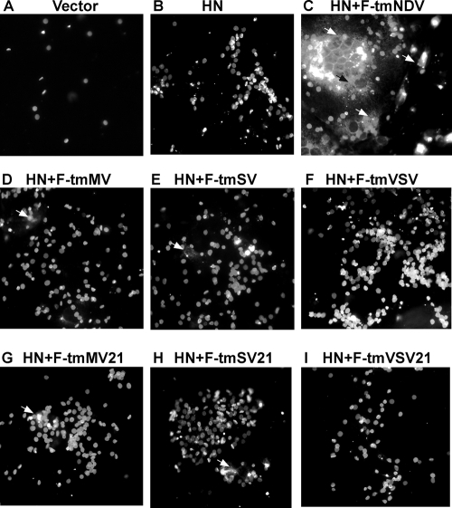
The role of specific sequences in the transmembrane (TM) domain of Newcastle disease virus (NDV) fusion (F) protein in the structure and function of this protein was assessed by replacing this domain with the F protein TM domains from two other paramyxoviruses, Sendai virus (SV) and measles virus (MV), or the TM domain of the unrelated glycoprotein (G) of vesicular stomatitis virus (VSV). Mutant proteins with the SV or MV F protein TM domains were expressed, transported to cell surfaces, and proteolytically cleaved at levels comparable to that of the wild-type protein, while mutant proteins with the VSV G protein TM domain were less efficiently expressed on cell surfaces and proteolytically cleaved. All mutant proteins were defective in all steps of membrane fusion, including hemifusion. In contrast to the wild-type protein, the mutant proteins did not form detectable complexes with the NDV hemagglutinin-neuraminidase (HN) protein. As determined by binding of conformation-sensitive antibodies, the conformations of the ectodomains of the mutant proteins were altered. These results show that the specific sequence of the TM domain of the NDV F protein is important for the conformation of the preactivation form of the ectodomain, the interactions of the protein with HN protein, and fusion activity.


Figures

FIG. 1.
FIG. 1.
Wild-type and mutant protein TM domains. The sequences of the wild-type NDV F protein (strain AV; F-tmNDV), MV F protein, SV F protein, and VSV G protein TMs and the junctions of the TM domains with the ectodomains and CT domains are shown at the top. The TM domain sequences of the six NDV F protein mutants are shown at the bottom. In the F-tmMV, F-tmSV, and F-tmVSV mutants, the 21-amino-acid NDV F protein TM domain was replaced with the 22-amino-acid TM domain of the MV F protein or the 23-amino-acid TM domain of the SV F or VSV G protein. In the F-tmMV21, F-tmSV21, and F-tmVSV21 mutants, the 21-amino-acid NDV F protein TM domain was replaced with the amino-terminal 21 amino acids from the TM domain of the MV F, SV F, or VSV G protein.
FIG. 2.
FIG. 2.
Expression of mutant F proteins. (A) Proteins in cell extracts of COS-7 cells transfected with cDNAs encoding wild-type or mutant F proteins or empty vector were resolved in 10% polyacrylamide gels under reducing conditions (top, +βME) or nonreducing conditions (bottom, −βME), and the F proteins were detected by Western blotting using anti-HR2 antibody. F0, uncleaved F protein; F1, the cleaved F protein; Fnr, nonreduced form of the protein (F0 and F1+F2). F proteins are defined in the legend to Fig. 1. (B) mRNAs generated by transcription of cDNAs encoding wild-type or mutant F proteins were translated in a rabbit reticulocyte cell-free system in the absence of membranes. The resulting radioactively labeled proteins were resolved on polyacrylamide gels and detected by autoradiography. Fug, unglycosylated F protein. (C) mRNAs generated by cell-free transcription were translated in a rabbit reticulocyte cell-free system containing dog pancreas membranes. Lanes 5 through 9 show products of reactions that also contained the peptide glycosylation inhibitor NYT. Fg, glycosylated F protein; Fug, unglycosylated F protein; F+ss, untranslocated F protein retaining the signal sequence; F-ss, translocated but unglycosylated F protein with signal sequence cleaved from the protein. Note that the order of proteins in panel B, lanes 1 through 5, and in panel C is different from the order in panels A and B, lanes 6 through 10.
FIG. 3.
FIG. 3.
Surface expression of wild-type and mutant F proteins. (A) Flow cytometry analyses of the levels of surface expression of three mutant proteins compared to that of the wild-type protein, detected by the binding of anti-NDV antibody after transfection of cells with cDNAs. Cells were processed for flow cytometry at 48 h posttransfection. Each panel shows the numbers of cells expressing the wild-type protein (dashed line), the background (cells transfected with vector and incubated with both primary and secondary antibodies; gray line), and cells expressing a mutant F protein (black line and arrow). (B) Surface expression of wild-type (F-tmNDV) or mutant F proteins (F-tmMV, F-tmSV, and F-tmVSV) detected by immunofluorescence using anti-NDV antibody. All images were acquired at 48 h posttransfection using the same exposures and were processed through Adobe Photoshop using identical settings for all images.
FIG. 4.
FIG. 4.
Hemifusion directed by mutant F proteins. Hemifusion was detected, as described in Materials and Methods and elsewhere in the text, by the spread of fluorescence-labeled lipid R18 from red blood cell membranes to underlying COS-7 cell monolayers. (A) Negative controls transfected with vector alone (pSVL). (B) Cells expressing HN protein alone. (C through I) Monolayers expressing HN protein with wild-type or mutant F proteins (as designated at the top of each panel) and bound fluorescent red blood cells. Positive fusion, present in panel C, is indicated by arrows. The panels show representative fields from multiple experiments. Rare hemifusion events in panels D, E, G, and H are indicated by arrows.
FIG. 5.
FIG. 5.
Syncytium and pore formation directed by mutant F proteins. (Left) Quantification of syncytium formation directed by wild-type or mutant F proteins in the presence of wild-type NDV HN protein. All proteins were expressed using the pSVL vector. Values are expressed as percentages of syncytium formation detected in cells transfected with wild-type F and HN proteins at 72 h posttransfection. Error bars indicate standard deviations. (Right) Quantification of content mixing directed by wild-type or mutant F proteins in the presence of wild-type HN protein as measured by β-galactosidase activities. Viral proteins were expressed using the pCAGGs vector. Values are expressed as percentages of content mixing detected in cells transfected with wild-type F and HN proteins. Error bars indicate standard deviations.
FIG. 6.
FIG. 6.
Virus specificity of coimmunoprecipitation of HN and F proteins. The cDNAs (plasmids) used for transfection are indicated at the bottoms of the panels. All cDNAs (NDV Fwt, F protein cDNA; HN, HN protein cDNA; NDV-F-FLAG, NDV F cDNA with the FLAG sequence inserted in-frame at the carboxyl terminus of the coding sequence; SV-F-FLAG, SV F cDNA with the FLAG sequence inserted in-frame at the carboxyl terminus of the coding sequence) were inserted into pSVL. (A) Surface expression of FLAG-tagged NDV F and SV F proteins. Surfaces of cells expressing these proteins were biotinylated as previously described (21, 22). After cell lysis, biotinylated molecules were precipitated with NeutrAvidin and the proteins in the precipitate were detected by anti-NDV F (anti-HR2) (left) or anti-FLAG (right) antibody. T, total proteins in cell extracts; B, biotinylated cell surface proteins. (B and C) Coimmunoprecipitation of HN protein with FLAG-tagged NDV F or SV F proteins. The antibodies used to precipitate proteins from each extract (IP) are indicated at the top of each lane in both panels. T is the total cell extract and represents 20% of that used in the immunoprecipitations. All proteins were electrophoresed in the presence of reducing agent. (B) Detection by Western blotting of NDV-F-FLAG or SV F-F-FLAG protein using a polyclonal antibody specific for the FLAG sequence (anti-FLAG). (C) Detection by Western blotting of HN protein in total extracts or in immunoprecipitates using anti-HN-AS antibody. WB, Western blot.
FIG. 7.
FIG. 7.
Coimmunoprecipitation of wild-type HN protein and mutant F proteins. Plasmids used for transfections are indicated at the bottoms of the lanes (all cDNAs were inserted into pSVL), and the antibodies used to precipitate proteins (IP) are indicated at the top of each lane. The anti-F protein antibody used for IP was anti-Fu1a. The anti-HN protein antibody used for IP was a mixture of anti-HN protein monoclonal antibodies. T is the total extract and represents 20% of the amount used in immunoprecipitations. Results shown are representative of at least two separate determinations for each mutant protein. (A) The F protein, detected in the Western blot (WB) using anti-F protein antibody (anti-HR2), was present in each precipitate and was electrophoresed in the absence of reducing agent. Results with HN and F-tmNDV controls were always accomplished in parallel and are shown. (B) HN protein present in each sample electrophoresed in the presence of reducing agent and detected in the Western blot using anti-HN (anti-HN-AS) protein antibody. Results with HN and F-tmNDV controls were always accomplished in parallel and are shown.
FIG. 8.
FIG. 8.
Binding of conformation-sensitive antibodies to surface-expressed mutant F proteins. (A) Binding of anti-Fu1a to cell surfaces. (Top) Representative immunofluorescence detected after binding anti-Fu1a to COS-7 cells expressing wild-type or mutant F proteins or empty vector (in the absence of HN protein expression). (Bottom) Graphs showing the quantification of these results and the results of identical experiments, accomplished as described in Materials and Methods. All images used for each data set were acquired using the same exposures and were processed through Adobe Photoshop using identical settings for all images. Error bars represent standard deviations. (B) Binding of anti-HR1 to cell surfaces expressing mutant proteins. (Top) Representative immunofluorescence detected after binding anti-HR1 antibody to COS-7 cells expressing wild-type or mutant F proteins or empty vector (in the absence of HN protein expression). (Bottom) Graphs showing the quantification of these results as well as those from identical experiments, accomplished as described in Materials and Methods. All images for each data set were acquired using the same exposures and were processed through Adobe Photoshop using identical settings for all images. Error bars represent standard deviations. The binding of anti-NDV antibody is quantified in Table 1.
FIG. 9.
FIG. 9.
Effect of HN protein expression on anti-HR1 binding to surface-expressed mutant F proteins. The panels show representative immunofluorescence detected after binding anti-HR1 antibody to COS-7 cells coexpressing wild-type or mutant F proteins and the HN protein. All images, acquired using the same exposures, were processed through Adobe Photoshop using identical settings for all images.

Similar articles

Cited by

References

    1. Aguilar, H. C., et al. 2007. Polybasic KKR motif in the cytoplasmic tail of Nipah virus fusion protein modulates membrane fusion by inside-out signaling. J. Virol. 81:4520-4532. - PMC - PubMed
    1. Aguilar, H. C., et al. 2006. N-glycans on Nipah virus fusion protein protect against neutralization but reduce membrane fusion and virus entry. J. Virol. 80:4878-4889. - PMC - PubMed
    1. Ali, A., and N. P. Nayak. 2000. Assembly of Sendai virus: M protein interacts with F and HN proteins and with the cytoplasmic tail and transmembrane domain of F protein. Virology 276:289-303. - PubMed
    1. Armstrong, R. T., A. S. Kushnir, and J. M. White. 2000. The transmembrane domain of influenza hemagglutinin exhibits a stringent length requirement to support the hemifusion to fusion transition. J. Cell Biol. 151:425-438. - PMC - PubMed
    1. Bagai, S., and R. A. Lamb. 1996. Truncation of the COOH-terminal region of paramyxovirus SV5 fusion protein leads to hemifusion but not complete fusion. J. Cell Biol. 135:73-84. - PMC - PubMed

Publication types

MeSH terms