. 2011 Feb;60(2):517-24.
doi: 10.2337/db10-0266.

Hyperglycemia activates caspase-1 and TXNIP-mediated IL-1beta transcription in human adipose tissue

Affiliations

Hyperglycemia activates caspase-1 and TXNIP-mediated IL-1beta transcription in human adipose tissue

Tim B Koenen et al. Diabetes. 2011 Feb.

Abstract

Objective: Obesity is characterized by elevated levels of proinflammatory cytokines, including interleukin (IL)-1β, that contribute to the development of insulin resistance. In this study, we set out to investigate whether hyperglycemia drives IL-1β production and caspase-1 activation in murine and human adipose tissue, thus inducing insulin resistance.

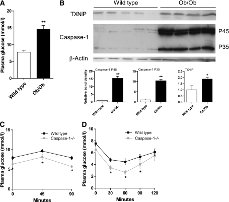
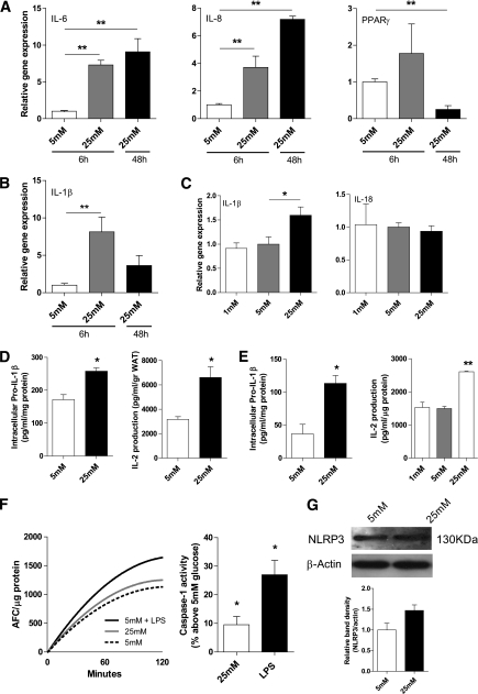
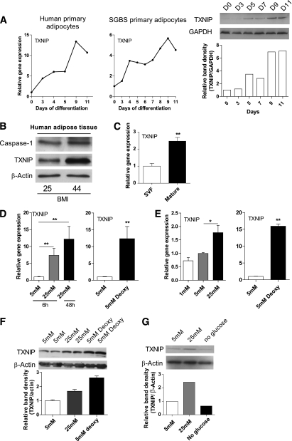
Research design and methods: ob/ob animals were used as a model to study obesity and hyperglycemia. Human adipose tissue fragments or adipocytes were cultured in medium containing normal or high glucose levels. Additionally, the role of thioredoxin interacting protein (TXNIP) in glucose-induced IL-1β production was assessed.

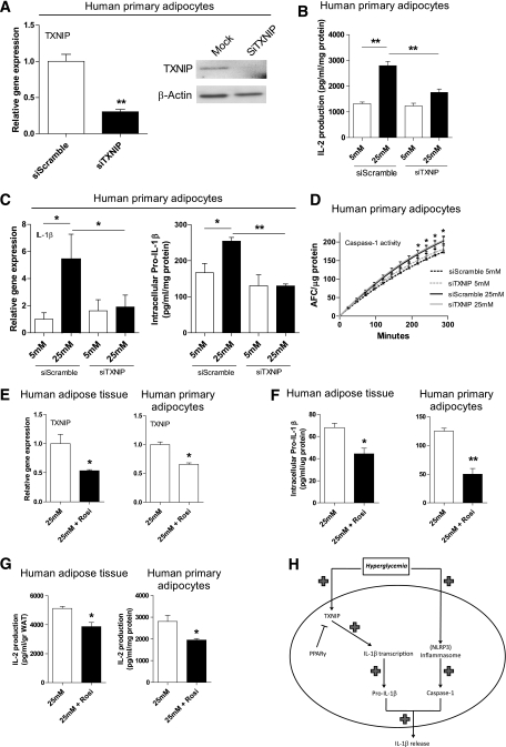
Results: TXNIP and caspase-1 protein levels were more abundantly expressed in adipose tissue of hyperglycemic ob/ob animals as compared with wild-type mice. In human adipose tissue, high glucose resulted in a 10-fold upregulation of TXNIP gene expression levels (P < 0.01) and a 10% elevation of caspase-1 activity (P < 0.05), together with induction of IL-1β transcription (twofold, P < 0.01) and a significant increase in IL-1β secretion. TXNIP suppression in human adipocytes, either by a small interfering RNA approach or a peroxisome proliferator-activated receptor-γ agonist, counteracted the effects of high glucose on bioactive IL-1 production (P < 0.01) mainly through a decrease in transcription levels paralleled by reduced intracellular pro-IL-1β levels.

Conclusions: High glucose activates caspase-1 in human and murine adipose tissue. Glucose-induced activation of TXNIP mediates IL-1β mRNA expression levels and intracellular pro-IL-1β accumulation in adipose tissue. The concerted actions lead to enhanced secretion of IL-1β in adipose tissue that may contribute to the development of insulin resistance.


Figures

FIG. 1.
FIG. 1.
TXNIP and caspase-1 protein levels are increased in the adipose tissue of ob/ob mice. A: Plasma glucose levels in fasted wild-type mice and ob/ob mice (n = 5 per group). B: Western blot images and quantification of TXNIP, procaspase-1 (p45), and active caspase-1 (p35) protein levels in the epididymal adipose tissue of wild-type mice and ob/ob mice (n = 5 per group). C: A glucose tolerance test was done using fasted wild-type and caspase-1−/− animals (n = 5 animals per group). D: An insulin tolerance test was performed in fasted wild-type and caspase-1−/− animals (n = 5 animals per group). *P < 0.05; **P < 0.01 using a Student t test.
FIG. 2.
FIG. 2.
Hyperglycemia induces proinflammatory gene expression and results in an increased production of IL-1 by intact adipose tissue and adipocytes. A: IL-6, IL-8, and PPAR-γ gene expression levels in human intact adipose tissue (n = 3) after 6 or 48 h of glucose treatment. B: IL-1β gene expression levels in human adipose tissue (n = 3) treated with glucose for 6 or 48 h. C: IL-1β and IL-18 gene expression levels in human primary adipocytes (n = 3) treated with various concentrations of glucose. D and E: Intracellular pro-IL-1β levels measured in lysates from intact human adipose tissue (D) or human primary adipocytes (E) treated with 5 or 25 mM glucose for 48 h (n = 4) and IL-2 production from NOB-1 cells after exposure to medium from intact human subcutaneous adipose tissue of three different donors treated with 5 or 25 mM of glucose for 48 h. F: Caspase-1 activity assay in primary human adipocytes treated with 5 mM, 25 mM of glucose, or LPS (10 ng/mL) for 48 h. The left graph displays the results of one representative experiment. The right graph displays the average results of n = 6 experiments. G: NLRP3 protein expression levels in human primary adipocytes treated with 5 or 25 mM of glucose for 48 h. A representative Western blot and quantification of the results (n = 3) are shown. *P < 0.05; **P value < 0.01 using a one-way ANOVA test (AC), Student t test (D and E), or a Wilcoxon rank test (F; right graph).
FIG. 3.
FIG. 3.
TXNIP is present in human adipose tissue and adipocytes and is regulated by glucose. A: TXNIP gene and protein expression levels during adipocyte differentiation of human primary adipocytes or human SGBS adipocytes. B: Western blot images of caspase-1 and TXNIP protein levels in total human subcutaneous adipose tissue (n = 2). C: Relative gene expression levels of TXNIP in mature adipocytes or the SVF isolated from human adipose tissue (n = 3). D: Gene expression levels of TXNIP in intact adipose tissue treated with various concentrations of glucose (n = 3). E: Gene expression levels of TXNIP in human primary adipocytes treated with various concentrations of glucose (n = 3). F: Protein levels of TXNIP in human adipose tissue after 5 mM glucose, 25 mM glucose, or 5 mM deoxyglucose treatment for 48 h (n = 2). G: Protein levels of TXNIP after glucose starvation (no glucose), 5 mM glucose, and 25 mM glucose for 48 h in human primary adipocytes. *P < 0.05; **P < 0.01 using a one-way ANOVA test (D and E) or a Student t test (C).
FIG. 4.
FIG. 4.
TXNIP reduction results in a decline of high glucose-induced IL-1 production by modulating IL-1β gene expression. A: Gene and protein expression levels of TXNIP after siRNA treatment against TXNIP in human primary adipocytes (n = 6). B: IL-2 production from NOB-1 cells after exposure to medium from human primary adipocytes transfected with TXNIP siRNA and treated with 5 or 25 mM glucose for 48 h (n = 6). C: IL-1β mRNA expression and intracellular pro-IL-1β levels in TXNIP siRNA treated adipocytes exposed to 5 or 25 mM glucose for 48 h (n = 4). D: Caspase-1 activity assay in TXNIP siRNA treated human primary adipocytes exposed to 5 or 25 mM glucose for 48 h (n = 3). E: TXNIP gene expression levels in intact human adipose tissue and primary adipocytes exposed to 25 mM glucose for 48 h with or without Rosiglitazone (10 μM) treatment for 24 h (n = 3). F: Intracellular pro-IL-1β levels measured in lysates of intact human adipose tissue and primary adipocytes exposed to 25 mM glucose for 48 h with or without Rosiglitazone (10 μM) treatment for 10 h (n = 3). G: IL-2 production from NOB-1 cells after exposure to medium from intact adipose tissue and human primary adipocytes treated with 25 mM glucose for 48 h with or without Rosiglitazone (10 μM) stimulation for 10 h (n = 4). H: Role of TXNIP in hyperglycemia-induced release of IL-1β from adipose tissue. *P < 0.05; **P < 0.01 using a one-way ANOVA test (B and C) or a Student t test (DG).

References

    1. Alexandraki K, Piperi C, Kalofoutis C, Singh J, Alaveras A, Kalofoutis A. Inflammatory process in type 2 diabetes: the role of cytokines. Ann N Y Acad Sci 2006;1084:89–117 - PubMed
    1. Katsuki A, Sumida Y, Murashima S, et al. Serum levels of tumor necrosis factor-alpha are increased in obese patients with noninsulin-dependent diabetes mellitus. J Clin Endocrinol Metab 1998;83:859–862 - PubMed
    1. Schenk S, Saberi M, Olefsky JM. Insulin sensitivity: modulation by nutrients and inflammation. J Clin Invest 2008;118:2992–3002 - PMC - PubMed
    1. Shoelson SE, Herrero L, Naaz A. Obesity, inflammation, and insulin resistance. Gastroenterology 2007;132:2169–2180 - PubMed
    1. Lin Y, Rajala MW, Berger JP, Moller DE, Barzilai N, Scherer PE. Hyperglycemia-induced production of acute phase reactants in adipose tissue. J Biol Chem 2001;276:42077–42083 - PubMed

Publication types

MeSH terms