. 2011 Feb 24;470(7335):498-502.
doi: 10.1038/nature09775. Epub 2011 Jan 26.

Transient Hoogsteen base pairs in canonical duplex DNA

Affiliations

Transient Hoogsteen base pairs in canonical duplex DNA

Evgenia N Nikolova et al. Nature. .

Abstract

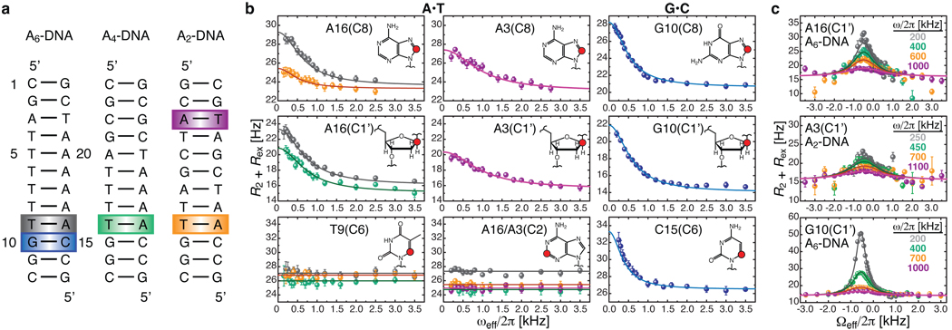
Sequence-directed variations in the canonical DNA double helix structure that retain Watson-Crick base-pairing have important roles in DNA recognition, topology and nucleosome positioning. By using nuclear magnetic resonance relaxation dispersion spectroscopy in concert with steered molecular dynamics simulations, we have observed transient sequence-specific excursions away from Watson-Crick base-pairing at CA and TA steps inside canonical duplex DNA towards low-populated and short-lived A•T and G•C Hoogsteen base pairs. The observation of Hoogsteen base pairs in DNA duplexes specifically bound to transcription factors and in damaged DNA sites implies that the DNA double helix intrinsically codes for excited state Hoogsteen base pairs as a means of expanding its structural complexity beyond that which can be achieved based on Watson-Crick base-pairing. The methods presented here provide a new route for characterizing transient low-populated nucleic acid structures, which we predict will be abundant in the genome and constitute a second transient layer of the genetic code.


Figures

Figure 1
Figure 1. Detection of base-pair specific excited states in CA/TG steps of duplex DNA
a, DNA constructs containing varying length A-tracts with color-coded A•T and G•C base-pairs at CA/TG steps that show carbon chemical exchange. b, On-resonance 13C R relaxation dispersion profiles for A•T (26.0 °C) and G•C (30.5 °C) showing CA/TG specific chemical exchange at purine base C8 and sugar C1’ and at cytosine base C6. Shown are the best base-pair global fits (solid line) to a two-state asymmetric exchange model (Supplementary Eq.1). c, Representative off-resonance relaxation dispersion profiles for corresponding C1’ sites and best global fits as in b. All error bars represent experimental uncertainty (one s.d.) estimated from mono-exponential fitting of duplicate sets of R data.
Figure 2
Figure 2. Kinetic-thermodynamic analysis of ground-to-excited state transitions
a, Representative on-resonance 13C R relaxation dispersion profiles as a function of temperature for A16 (A6-DNA and A4-DNA), A3 (A2-DNA), and G10 (A6-DNA) C1’. b, Modified van’t Hoff plots showing temperature dependence of the forward (kA) and reverse (kB) rate constants for the two-site exchange in A•T and G•C base-pairs highlighted in Fig. 1a. Error bars represent experimental uncertainty (one s.d.) as determined from propagation of errors obtained from mono-exponential fitting of duplicate sets of R data. c, Corresponding kinetic-thermodynamic profiles for exchange between the Watson-Crick (WC) ground state and the excited state (ES) via a transition state (‡), showing activation and net free energy (G), enthalpy (H), and entropy (TS) changes (referenced to 0).
Figure 3
Figure 3. Chemical shift assignment of excited state Hoogsteen base-pairs
a, Chemical structures for WC and HG A•T and G•C+ base-pairs. The HG geometry can be achieved by purine rotation around the glycosidic bond (χ) and base-flipping (θ), affecting simultaneously C8 and C1’ (yellow). b, Depiction of C8 and C1’ chemical shifts relative to WC for the excited state (ES, grey); an N1-methyladenine modified A6-DNA (HG(1mA), red), an N1-methylguanine modified A6-DNA (HG(1mG), violet), and an echinomycin-bound DNA (HG(drug), green) forming HG base-pairs; a simulated A16•T9 HG base-pair in A6-DNA (HG(MD), blue); a C3’-endo locked A6-DNA (WC(LNA), orange); and representative cartoons.
Figure 4
Figure 4. Watson-Crick to Hoogsteen base-pair transition simulations
a, Pseudo-free-energy (E, kcal/mol) contour plots as a function of (χ, θ) pairs for A16•T9 obtained from multiple CPR trajectories (a–d). b, Initial WC and final exited-state (ES) HG structures and representative lowest-energy (b1) and highest-energy (c3) transition state (‡) structures of A16•T9 illustrating the span of CPR transition barriers, and their relative potential energies (averaged for WC and HG) compared with enthalpies (H) derived from NMR R relaxation dispersion for chemical exchange or NMR imino proton exchange for A•T base-pair opening. c, Snapshots from a representative CPR transition pathway (a1) for A16•T9.

Comment in

References

    1. Watson JD, Crick FH. Molecular structure of nucleic acids; a structure for deoxyribose nucleic acid. Nature. 1953;171:737–738. - PubMed
    1. Record MT, Jr, et al. Double helical DNA: conformations, physical properties, and interactions with ligands. Annu Rev Biochem. 1981;50:997–1024. - PubMed
    1. Koudelka GB, Mauro SA, Ciubotaru M. Indirect readout of DNA sequence by proteins: the roles of DNA sequence-dependent intrinsic and extrinsic forces. Prog Nucleic Acid Res Mol Biol. 2006;81:143–177. - PubMed
    1. Rohs R, et al. The role of DNA shape in protein-DNA recognition. Nature. 2009;461:1248–1253. - PMC - PubMed
    1. Segal E, et al. A genomic code for nucleosome positioning. Nature. 2006;442:772–778. - PMC - PubMed

Publication types