. 2011 May;13(5):1205-15.
doi: 10.1111/j.1462-2920.2010.02420.x. Epub 2011 Jan 27.

Demethylation of methylarsonic acid by a microbial community

Affiliations

Demethylation of methylarsonic acid by a microbial community

Masafumi Yoshinaga et al. Environ Microbiol. 2011 May.

Abstract

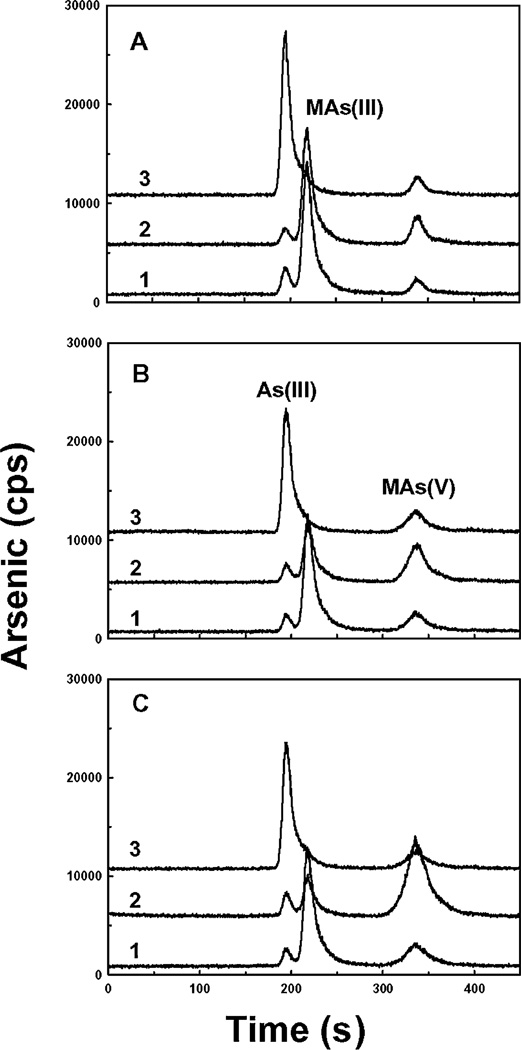
Arsenic is one of the most widespread environmental carcinogens and has created devastating human health problems worldwide, yet little is known about mechanisms of biotransformation in contaminated regions. Methylarsonic acid [MAs(V)], extensively utilized as an herbicide, is largely demethylated to more toxic inorganic arsenite, which causes environmental problems. To understand the process of demethylation of methylarsenicals, soil samples commonly used on Florida golf courses were studied. Several soil extracts were found to demethylate MAs(V) to inorganic arsenite [As(III)]. From these extracts, a bacterial isolate was capable of reducing MAs(V) to MAs(III) but not of demethylating to As(III). A second bacterial isolate was capable of demethylating MAs(III) to As(III) but not of reducing MAs(V). A mixed culture could carry out the complete process of reduction and demethylation, demonstrating that demethylation of MAs(V) to As(III) is a two-step process. Analysis of the 16S ribosomal DNA sequences of the two organisms identified the MAs(V)-reducing and the MAs(III)-demethylating isolates as belong to Burkholderia and Streptomyces species respectively. This is the first report of a novel pathway of degradation of a methylarsenical herbicide by sequential reduction and demethylation in a microbial soil community, which we propose plays a significant role in the arsenic biogeocycle.


Figures

Fig. 1
Fig. 1
Demethylation of MAs(V) to As(III) by soil extracts. A. Transformation of MAs(V) by soil extracts was analysed after 1 week incubation at room temperature by reverse-phase HPLC-ICP-MS, as described under Experimental procedures, where the x-axis represents retention time, and the y-axis represents relative amounts of arsenic expressed as counts per second (cps). Curve 1: 1 µM MAs(V) in ST 10−1 medium. Curve 2: S+P extracted in ST 10−1 medium. Curve 3: autoclaved S+P extracted in ST 10−1 medium containing 1 µM MAs(V). Curve 4: S extracted in ST 10−1 medium containing 1 µM MAs(V). Curve 5: S+P extracted in ST 10−1 medium containing 1 µM MAs(V). B. The transformation of 1 µM MAs(V) by S+P soil extracted in ST 10−1 medium was assayed after incubation for 3 (curve 2), 5 (curve 3) and 7 (curve 4) days at room temperature. Curve 1: 1 µM MAs(V) control incubated in ST 10−1 medium for 7 days.
Fig. 2
Fig. 2
Reduction of MAs(V) by Burkholderia sp. MR1. Reduction of MAs(V) by a bacterial isolate identified as Burkholderia sp. MR1 was assayed by reverse-phase HPLC-ICP-MS, as described under Experimental procedures. Curve 1: 1 µM MAs(V) in ST 10−1 medium. Curve 2: cells of Burkholderia sp. MR1 in ST 10−1 medium containing 1 µM MAs(V).
Fig. 3
Fig. 3
Effect of growth media on MAs(V) reductase activity of Burkholderia sp. MR1 and P. putida KT2440. A–C. Each bacterial strain was inoculated into ST 10−1 medium (A), LB medium (B) or M9 medium supplemented with 0.2% glucose as carbon source (C), and incubated with 1 µM MAs(V) for 3 days at room temperature, following which reduction was analysed by reverse-phase HPCL-ICP-MS. D–F. Each strain was cultured overnight in LB medium, washed and suspended at a density of A600 of 3.0 in ST 10−1 medium (A), LB medium (B) or M9 medium supplemented with 0.2% glucose as carbon source (C), and then incubated with 1 µM MAs(V) at room temperature for 6 h. In each panel curve 1 shows medium with no cells; curve 2 shows P. putida KT2440 and curve 3 shows Burkholderia sp. MR1.
Fig. 4
Fig. 4
Uptake of MAs(V). The ability of Burkholideria sp. MR1 and P. putida KT2440 to take up MAs(V) was assayed as described under Experimental procedures. (●), Burkholderia sp. MR1 in ST 10−1 medium; (○), Burkholderia sp. MR1 in LB medium; (∆), P. pudita KT2440 in ST 10−1 medium; (▼), P. pudita KT2440 in LB medium. The error bars represent the standard deviation of three assays.
Fig. 5
Fig. 5
The P. putida ars operon does not encode a MAs(V) reductase. MAs(V) reduction was assayed in the indicated bacterial cultures following growth in ST 10−1 medium containing 1 µM MAs(V) for 3 days at room temperature. Curve 1: 1 µM MAs(V) control in ST 10−1 medium; curve 2: E. coli strain W3110; curve 3: Burkholderia sp. MR1; curve 4: P. pudita strain Tec1; curve 5: P. putida strain ∆Ars1&2.
Fig. 6
Fig. 6
Demethylation of MAs(V) by Streptomyces sp. MD1. Demethylation of MAs(III) by a bacterial isolate identified as Streptomyces sp. MD1 was assayed by reverse-phase HPLC-ICP-MS, as described under Experimental procedures. A. Demethylation of MAs(V) in mixed culture. Strains were incubated separately or together in filter-sterilized S+P extract containing 1 µM MAs(V) for 1 week at room temperature. Curve 1: 1 µM MAs(V) in filter-sterilized S+P. Curve 2: Burkholderia sp. MR1. Curve 3: Streptomyces sp. MD1. Curve 4: co-culture of Burkholderia sp. MR1 and Streptomyces sp. MD1. B. Demethylation of MAs(III) by Streptomyces sp. MD1. MAs(III) was prepared by chemical reduction of MAs(V), as described under Experimental procedures. Line 1: MAs(III) in filter sterilized S+P extract without incubation. Line 2: MAs(III) in filter-sterilized S+P extract after three days of incubation at room temperature. Line 3: Streptomyces sp. MD1 incubated with MAs(III) for 3 days at room temperature.
Fig. 7
Fig. 7
Effect of growth media on MAs(III) demethylating activity of Streptomyces sp. MD1. Streptomyces sp. MD1 was grown overnight in LB medium at 37°C, washed and suspended the same volume in ST 10−1 medium (A), LB medium (B) or M9 medium supplemented with 0.2% glucose as carbon source (C), and incubated with 1 µM MAs(III) for 3 h at room temperature, following which demethylation was analysed by reverse-phase HPCL-ICP-MS. In each panel curve 1 shows 1 µM MAs(III) in medium with no cells and no incubation; curve 2 shows 1 µM MAs(III) in medium with no cells after 3 h; and curve 3 shows Streptomyces sp. MD1 incubated for 3 h in medium containing 1 µM MAs(III).
Fig. 8
Fig. 8
Demethylation of MAs(V) is a two-step process carried out by a microbial community. Demethylation of MAs(V) in soil takes place by sequential reduction to MAs(III) by Burkholderia sp. MR1 followed by demethylation of MAs(III) to As(III) by Streptomyces sp. MD1. Burkholderia sp. MR1 takes up MAs(V) by unknown transporters (a), possibly phosphate transporters or AQPs, and extrudes MAs(III) by efflux systems (b), possibly ArsB, Acr3 or AQPs. Streptomyces sp. MD1 takes up MAs(III), possibly by AQPs or glucose permeases (c), and extrudes As(III) by ArsB or Acr3 (d).

References

    1. Abernathy CO, Liu YP, Longfellow D, Aposhian HV, Beck B, Fowler B, et al. Arsenic: health effects, mechanisms of actions, and research issues. Environ Health Perspect. 1999;107:593–597. - PMC - PubMed
    1. Akkari KH, Frans RE, Lavy TL. Factors affecting degradation of MSMA in soil. Weed Sci. 1986;34:781–788.
    1. Bachmann BJ. In: Derivations and genotypes of some mutant derivatives of Escherichia coli K12. In Escherichia coli and Salmonella typhimurium: Cellular and Molecular Biology. Neidhardt FC, Ingraham JL, Low KB, Magasanik B, Schaechter M, Umbarger HE, editors. Washington, DC, USA: American Society for Microbiology; 1987. pp. 1190–1219.
    1. Bentley R, Chasteen TG. Microbial methylation of metalloids: arsenic, antimony, and bismuth. Microbiol Mol Biol Rev. 2002;66:250–271. - PMC - PubMed
    1. Bhattacharjee H, Rosen BP. Arsenic metabolism in prokaryotic and eukaryotic microbes. In: Nies DHS, Simon S, editors. Molecular Microbiology of Heavy Metals. Heidelberg, Germany: Springer-Verlag; 2007. pp. 371–406.

Publication types