

Differential patterns of histone acetylation in inflammatory bowel diseases
- PMID: 21272292
- PMCID: PMC3040698
- DOI: 10.1186/1476-9255-8-1
Differential patterns of histone acetylation in inflammatory bowel diseases
Abstract
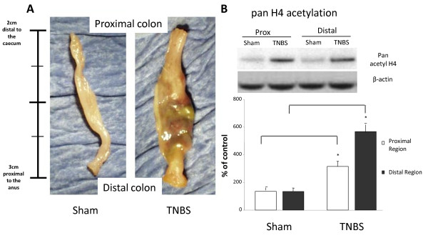
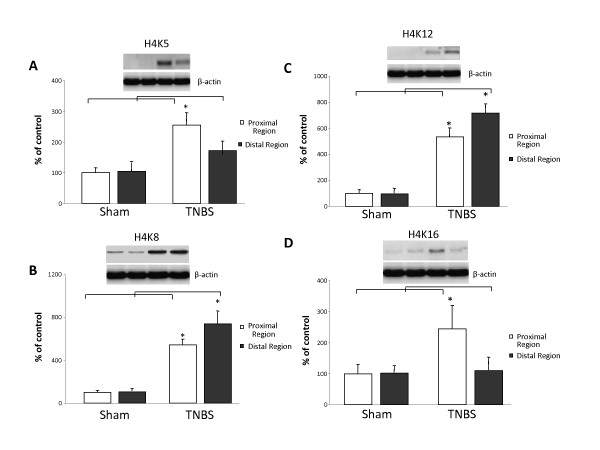
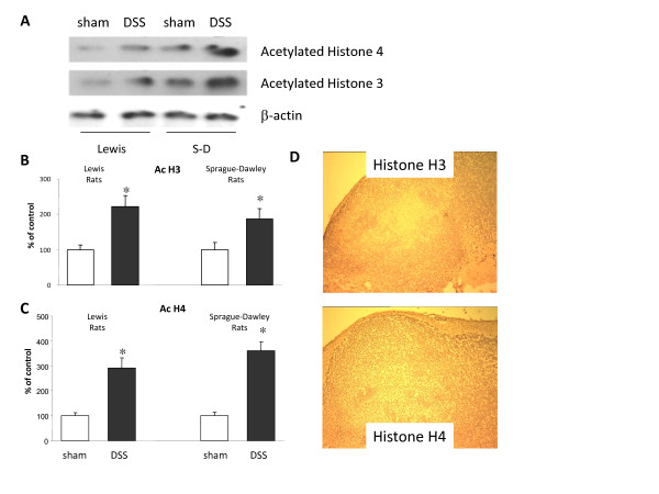
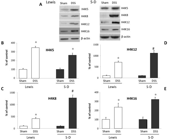
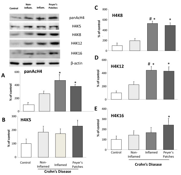
Post-translational modifications of histones, particularly acetylation, are associated with the regulation of inflammatory gene expression. We used two animal models of inflammation of the bowel and biopsy samples from patients with Crohn's disease (CD) to study the expression of acetylated histones (H) 3 and 4 in inflamed mucosa. Acetylation of histone H4 was significantly elevated in the inflamed mucosa in the trinitrobenzene sulfonic acid model of colitis particularly on lysine residues (K) 8 and 12 in contrast to non-inflamed tissue. In addition, acetylated H4 was localised to inflamed tissue and to Peyer's patches (PP) in dextran sulfate sodium (DSS)-treated rat models. Within the PP, H3 acetylation was detected in the mantle zone whereas H4 acetylation was seen in both the periphery and the germinal centre. Finally, acetylation of H4 was significantly upregulated in inflamed biopsies and PP from patients with CD. Enhanced acetylation of H4K5 and K16 was seen in the PP. These results demonstrate that histone acetylation is associated with inflammation and may provide a novel therapeutic target for mucosal inflammation.
Figures
Similar articles
-
Nano-electrospray tandem mass spectrometric analysis of the acetylation state of histones H3 and H4 in stationary phase in Saccharomyces cerevisiae.BMC Biochem. 2011 Jul 4;12:34. doi: 10.1186/1471-2091-12-34. BMC Biochem. 2011. PMID: 21726436 Free PMC article.
-
Mass spectrometry analysis of the variants of histone H3 and H4 of soybean and their post-translational modifications.BMC Plant Biol. 2009 Jul 31;9:98. doi: 10.1186/1471-2229-9-98. BMC Plant Biol. 2009. PMID: 19643030 Free PMC article.
-
Impact of resuscitation strategies on the acetylation status of cardiac histones in a swine model of hemorrhage.Resuscitation. 2008 Feb;76(2):299-310. doi: 10.1016/j.resuscitation.2007.07.030. Epub 2007 Sep 5. Resuscitation. 2008. PMID: 17822827
-
Pleiotropic effects of the histone deacetylase Hos2 linked to H4-K16 deacetylation, H3-K56 acetylation, and H2A-S129 phosphorylation in Beauveria bassiana.Cell Microbiol. 2018 Jul;20(7):e12839. doi: 10.1111/cmi.12839. Epub 2018 Apr 6. Cell Microbiol. 2018. PMID: 29543404
-
Induction by fructose force-feeding of histone H3 and H4 acetylation at their lysine residues around the Slc2a5 gene and its expression in mice.Biosci Biotechnol Biochem. 2013;77(11):2188-91. doi: 10.1271/bbb.130300. Epub 2013 Nov 7. Biosci Biotechnol Biochem. 2013. PMID: 24200777
Cited by
-
Microbiota-sensitive epigenetic signature predicts inflammation in Crohn's disease.JCI Insight. 2018 Sep 20;3(18):e122104. doi: 10.1172/jci.insight.122104. eCollection 2018 Sep 20. JCI Insight. 2018. PMID: 30232290 Free PMC article.
-
The role of epigenetic modifications for the pathogenesis of Crohn's disease.Clin Epigenetics. 2021 May 12;13(1):108. doi: 10.1186/s13148-021-01089-3. Clin Epigenetics. 2021. PMID: 33980294 Free PMC article. Review.
-
STAT1 epigenetically regulates LCP2 and TNFAIP2 by recruiting EP300 to contribute to the pathogenesis of inflammatory bowel disease.Clin Epigenetics. 2021 Jun 10;13(1):127. doi: 10.1186/s13148-021-01101-w. Clin Epigenetics. 2021. PMID: 34112215 Free PMC article.
-
New Insights Into the Epigenetic Regulation of Inflammatory Bowel Disease.Front Pharmacol. 2022 Jan 31;13:813659. doi: 10.3389/fphar.2022.813659. eCollection 2022. Front Pharmacol. 2022. PMID: 35173618 Free PMC article. Review.
-
Short-term tissue permeability actions of dextran sulfate sodium studied in a colon organ culture system.Tissue Barriers. 2020 Apr 2;8(2):1728165. doi: 10.1080/21688370.2020.1728165. Epub 2020 Feb 20. Tissue Barriers. 2020. PMID: 32079482 Free PMC article.
References
-
- D'Haens G, Daperno M. Advances in biologic therapy for ulcerative colitis and Crohn's disease. Curr Gastroenterol. 2006;8(6):506–512. - PubMed
Grants and funding
LinkOut - more resources
Full Text Sources
Research Materials
Miscellaneous
