. 2011 Jan 27;2(1):4.
doi: 10.1186/scrt45.

Fetal derived embryonic-like stem cells improve healing in a large animal flexor tendonitis model

Affiliations

Fetal derived embryonic-like stem cells improve healing in a large animal flexor tendonitis model

Ashlee E Watts et al. Stem Cell Res Ther. .

Abstract

Introduction: Tendon injury is a common problem in athletes, with poor tissue regeneration and a high rate of re-injury. Stem cell therapy is an attractive treatment modality as it may induce tissue regeneration rather than tissue repair. Currently, there are no reports on the use of pluripotent cells in a large animal tendon model in vivo. We report the use of intra-lesional injection of male, fetal derived embryonic-like stem cells (fdESC) that express Oct-4, Nanog, SSEA4, Tra 1-60, Tra 1-81 and telomerase.

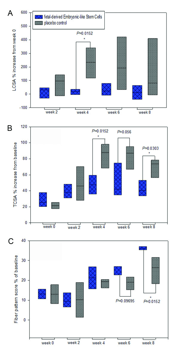
Methods: Tendon injury was induced using a collagenase gel-physical defect model in the mid-metacarpal region of the superficial digital flexor tendon (SDFT) of eight female adult Thoroughbred or Thoroughbred cross horses. Tendon lesions were treated one week later with intra-lesional injection of male derived fdESCs in media or media alone. Therapy was blinded and randomized. Serial ultrasound examinations were performed and final analysis at eight weeks included magnetic resonance imaging (MRI), biochemical assays (total DNA, glycosaminoglycan, collagen), gene expression (TNC, TNMD, SCX, COL1A1, COL3A1, COMP, DCN, MMP1, MMP3, MMP13, 18S) and histology. Differences between groups were assessed with Wilcoxon's rank sum test.

Results: Cell survival was demonstrated via the presence of the SRY gene in fdESC treated, but not control treated, female SDFT at the end of the trial. There were no differences in tendon matrix specific gene expression or total proteoglycan, collagen or DNA of tendon lesions between groups. Tissue architecture, tendon size, tendon lesion size, and tendon linear fiber pattern were significantly improved on histologic sections and ultrasound in the fdESC treated tendons.

Conclusions: Such profound structural effects lend further support to the notion that pluripotent stem cells can effect musculoskeletal regeneration, rather than repair, even without in vitro lineage specific differentiation. Further investigation into the safety of pluripotent cellular therapy as well as the mechanisms by which repair was improved seem warranted.


Figures

Figure 1
Figure 1
Study timeline. CONT, placebo control; fdESC, fetal derived embryonic-like stem cells; MRI, magnetic resonance imaging; U/S, ultrasound.
Figure 2
Figure 2
Ultrasound measurements. Normalized (A) lesion and (B) tendon cross-sectional area (CSA) and (C) linear fiber pattern score for fetal-derived Embryonic-like Stem Cells (fdESC) and placebo control (CONT) treated tendons at time points post treatment injection in weeks. Asterisks mark significantly lower (A, B) or significantly higher (C) values for fdESC than CONT treated tendons (one-tailed P < 0.05).
Figure 3
Figure 3
Transverse ultrasound images. Images were made 16 cm distal to the accessory carpal bone, eight weeks post treatment with A) fetal-derived Embryonic-like Stem Cells or B) placebo control injections. Lateral is to the right. Dotted lines outline the superficial digital flexor tendon and lesion. Arrowheads identify remaining treatment injection needle tracts.
Figure 4
Figure 4
Transverse T1 MR images. Images were made at 16 cm distal to the accessory carpal bone, post mortem, eight weeks after treatment injection with A) fetal derived Embryonic-like Stem Cells and B) placebo control. Lateral is to the right. Arrow-heads outline the treated tendon in the first image of each group.
Figure 5
Figure 5
Longitudinal histology. This figure shows 50× and 200× magnification of longitudinal sections of superficial digital flexor tendon stained with H&E and Picrosirius Red for A) fetal-derived Embryonic-like stem cell treated tendon and B) placebo control treated tendon. Picrosirius images shown under polarized light. Bars = 200 μm.
Figure 6
Figure 6
In situ hybridization longitudinal histology. 400× magnification of in-situ hybridization against genomic SRY in A) fetal-derived Embryonic-like stem cell treated tendon and B) placebo control treated tendon. Bar = 50 μm.

References

    1. Sharma P, Maffulli N. Biology of tendon injury: healing, modeling and remodeling. J Musculoskelet Neuronal Interact. 2006;6:181–90. - PubMed
    1. Kader D, Saxena A, Movin T, Maffulli N. Achilles tendinopathy: some aspects of basic science and clinical management. Br J Sports Med. 2002;36:239–249. doi: 10.1136/bjsm.36.4.239. - DOI - PMC - PubMed
    1. Jeffcott LB, Rossdale PD, Freestone J, Frank CJ, Towers-Clark PF. An assessment of wastage in thoroughbred racing from conception to 4 years of age. Equine Vet J. 1982;14:185–198. doi: 10.1111/j.2042-3306.1982.tb02389.x. - DOI - PubMed
    1. Rossdale PD, Hopes R, Digby NJ, Offord K. Epidemiological study of wastage among racehorses 1982 and 1983. Vet Rec. 1985;116:66–69. doi: 10.1136/vr.116.3.66. - DOI - PubMed
    1. Abate M, Gravare Silbernagel K, Siljeholm C, Di Iorio A, De Amicis D, Salini V, Werner S, Paganelli R. Pathogenesis of tendinopathies: inflammation or degeneration? Arthritis Res Ther. 2009;11:235. doi: 10.1186/ar2723. - DOI - PMC - PubMed

Publication types

MeSH terms