

Malignant PEComa: a case report with emphasis on clinical and morphological criteria
- PMID: 21272348
- PMCID: PMC3042371
- DOI: 10.1186/1471-2482-11-3
Malignant PEComa: a case report with emphasis on clinical and morphological criteria
Abstract
Background: Malignant perivascular epitheliod cell tumor (PEComa) is a very rare entity composed of distinctive perivascular epitheliod cells with variable immunoreactivity for melanocytic and muscle markers. At present this neoplasm does not have a known normal cellular counterpart and the natural history is often unpredictable. Up to now, few cases of PEComa have been described and treatment modalities are still controversial, particularly in advanced conditions.
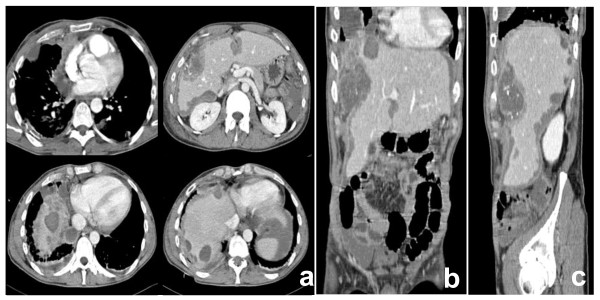
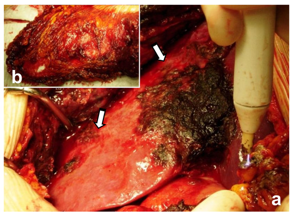
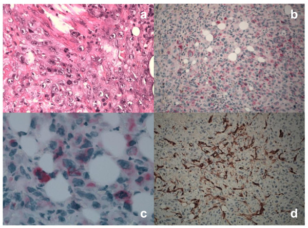
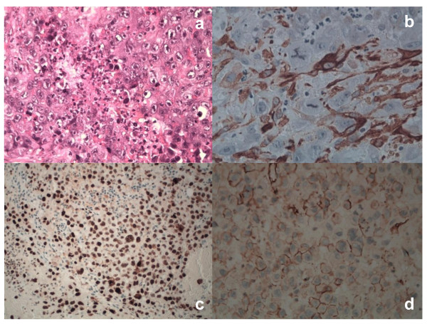
Case presentation: We handled the case of a 42-year-old man with unresectable PEComa of the abdomen. A 7 cm hepatic hypodense lesion between segment V and VIII of the liver and diffuse intraperitoneal nodules of 0,3-3,5 cm along the right subcapsular hepatic region, were documented by a CT scan. Radiological images showed abnormal lymph nodes of the right internal mammary chain and anterior mediastinum. The patient underwent an explorative laparotomy for uncontrolled intraabdominal hemorrhage without a well-defined preoperative tumor diagnosis. At surgery, multiple lobulated nodules containing hemorrhagic fluid on the liver surface, peritoneum and omentum were confirmed. The procedure had a palliative intent and consisted of hemostasis, hematomas evacuation and omentectomy. The diagnosis of PEComa was made after surgery on the basis of morphological and immunohistochemical criteria. Radiological and intra operative findings suggest that the mass has an hepatic origin with diffuse involvement of hepatic capsule and suspensory ligaments. The patient received medical support care with blood and plasma transfusions. In our experience, PEComa was clinically malignant, leading to a fatal outcome 25 days after hospital admission of patient.
Conclusions: Here we report and discuss the peculiar clinical, radiological and morphological presentation of unresectable PEComa. Although in the majority of the reported series, PEComas show a more better prognosis, our case presents with a particular aggressive biological behaviour. The importance of a correct preoperative diagnosis, the need for more effective targeted therapies based on tumor molecular knowledge and evidence-based clinical studies are emphasized together with a revision of the concerning scientific literature.
Figures
References
-
- Priola AM, Priola SM, Cataldi A, Marci V, Fava C. Acute abdomen as an unusual presentation of hepatic PEComa. A case report. Tumori. 2009;95:123–128. - PubMed
-
- Martignoni G, Pea M, Reghellin D, Gobbo S, Zamboni G, Chilosi M, Bonetti F. Molecular pathology of lymphangioleiomyomatosis and other perivascular epithelioid cell tumors. Arch Pathol Lab Med. 2010;134:33–40. - PubMed
-
- Baek JH, Chung MG, Jung DH, Oh JH. Perivascular epithelioid cell tumor (PEComa) in the transverse colon of an adolescent: a case report. Tumori. 2007;93:106–108. - PubMed
Publication types
MeSH terms
LinkOut - more resources
Full Text Sources
Other Literature Sources
