Comparative Study
. 2011 Jan 27;8(1):6.
doi: 10.1186/1743-8977-8-6.

Inflammatory and cytotoxic responses of an alveolar-capillary coculture model to silica nanoparticles: comparison with conventional monocultures

Affiliations
Comparative Study

Inflammatory and cytotoxic responses of an alveolar-capillary coculture model to silica nanoparticles: comparison with conventional monocultures

Jennifer Kasper et al. Part Fibre Toxicol. .

Abstract

Background: To date silica nanoparticles (SNPs) play an important role in modern technology and nanomedicine. SNPs are present in various materials (tyres, electrical and thermal insulation material, photovoltaic facilities). They are also used in products that are directly exposed to humans such as cosmetics or toothpaste. For that reason it is of great concern to evaluate the possible hazards of these engineered particles for human health. Attention should primarily be focussed on SNP effects on biological barriers. Accidentally released SNP could, for example, encounter the alveolar-capillary barrier by inhalation. In this study we examined the inflammatory and cytotoxic responses of monodisperse amorphous silica nanoparticles (aSNPs) of 30 nm in size on an in vitro coculture model mimicking the alveolar-capillary barrier and compared these to conventional monocultures.

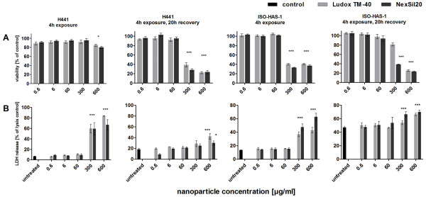
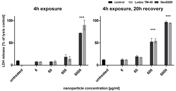
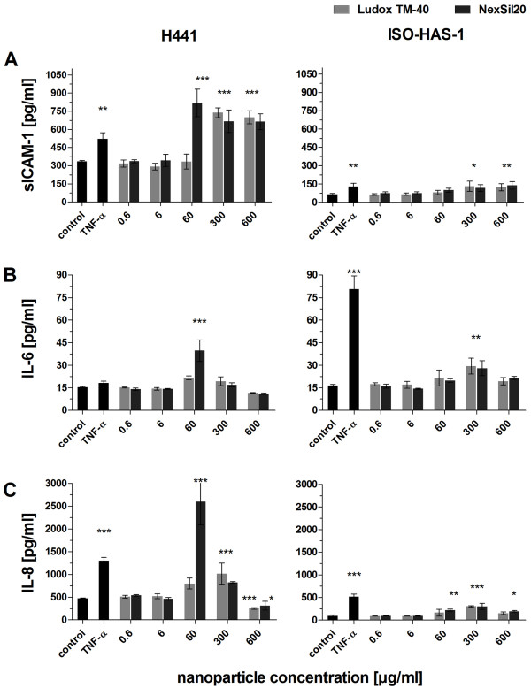
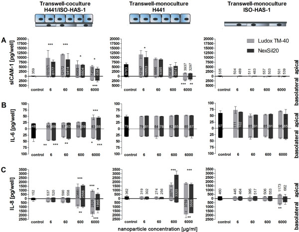
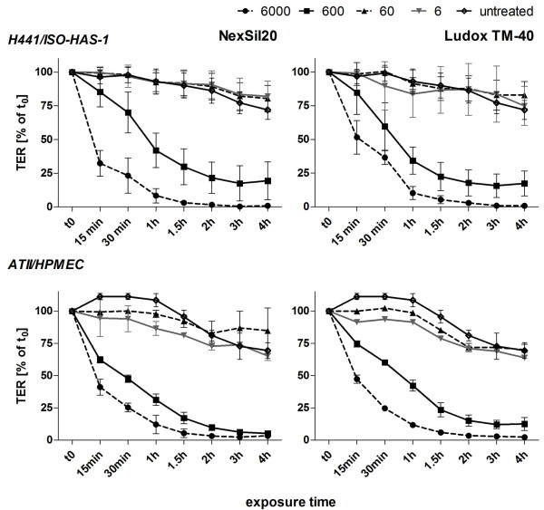
Methods: Thus, the epithelial cell line, H441, and the endothelial cell line, ISO-HAS-1, were used in monoculture and in coculture on opposite sides of a filter membrane. Cytotoxicity was evaluated by the MTS assay, detection of membrane integrity (LDH release), and TER (Transepithelial Electrical Resistance) measurement. Additionally, parameters of inflammation (sICAM-1, IL-6 and IL-8 release) and apoptosis markers were investigated.

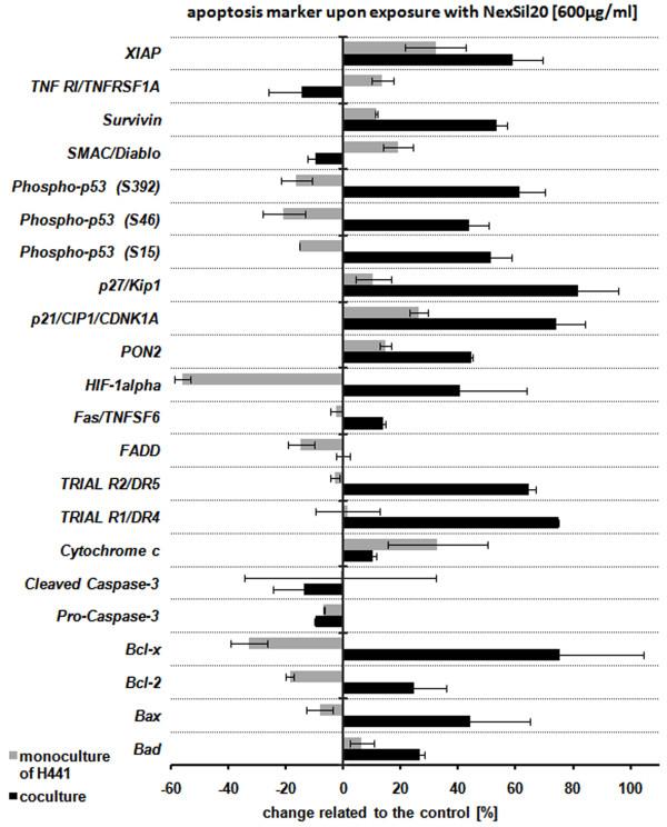
Results: Regarding toxic effects (viability, membrane integrity, TER) the coculture model was less sensitive to apical aSNP exposure than the conventional monocultures of the appropriate cells. On the other hand, the in vitro coculture model responded with the release of inflammatory markers in a much more sensitive fashion than the conventional monoculture. At concentrations that were 10-100fold less than the toxic concentrations the apically exposed coculture showed a release of IL-6 and IL-8 to the basolateral side. This may mimic the early inflammatory events that take place in the pulmonary alveoli after aSNP inhalation. Furthermore, a number of apoptosis markers belonging to the intrinsic pathway were upregulated in the coculture following aSNP treatment. Analysis of the individual markers indicated that the cells suffered from DNA damage, hypoxia and ER-stress.

Conclusion: We present evidence that our in vitro coculture model of the alveolar-capillary barrier is clearly advantageous compared to conventional monocultures in evaluating the extent of damage caused by hazardous material encountering the principle biological barrier in the lower respiratory tract.


Figures

Figure 1
Figure 1
Mitochondrial activity was measured via the MTS assay (A) and membrane integrity was determined by the LDH assay (B) for monocultures of H441 and ISO-HAS-1 on 96 well plates (conventional monoculture). Cells were incubated with aSNP (Ludox TM-40: light grey, NexSil20: dark grey) for 4 h in serum-free medium. aSNPs were then removed and cells were cultivated for further 20 h. The assays were conducted after both time points (4 h exposure and 4 h exposure with 20 h recovery). Data are depicted as percentage of the untreated control (A: MTS) or as percentage of the total LDH amount of the cells (B: LDH, lysis control). Results are shown as means ± S.D. (n = 6-9) of 2-3 independent experiments. *P < 0.05, **P < 0.01 and ***P < 0.001 compared to the untreated control
Figure 2
Figure 2
Membrane integrity was determined by the LDH assay for H441 in coculture. The H441 of the coculture were incubated with aSNP (Ludox TM-40: light grey, NexSil20: dark grey) for 4 h in serum-free medium. aSNPs were then removed and cells were cultivated for further 20 h. The assays were conducted after both time points (4 h exposure and 4 h exposure with 20 h recovery). Data are depicted as percentage of the total LDH amount of the cells (lysis control). Results are shown as means ± S.D. (n = 6-9) of 2-3 independent experiments. *P < 0.05, **P < 0.01 and ***P < 0.001 compared to the untreated control
Figure 3
Figure 3
The release of inflammatory mediators was measured after aSNP (Ludox TM-40: light grey, NexSil20: dark grey) exposure of monocultures of H441 and ISO-HAS-1 on 96 well plates (conventional monoculture). After 4 h incubation, aSNPs were removed and the cells were cultivated for further 20 h to detect sICAM-1, IL-6 and IL-8 release. Data are depicted as means ± S.D. of one representative experiment out of three independent experiments with n = 3 samples for each treatment. All independent experiments showed a comparable reaction following aSNP treatment. *P < 0.05, ** P < 0.01 and *** P < 0.001 compared to the untreated control
Figure 4
Figure 4
The release of inflammatory mediators (sICAM-1, IL-6 and IL-8) is shown after aSNP (Ludox TM-40: light grey, NexSil20: dark grey) incubation on apical/basolateral differentiated cocultures of H441 and ISO-HAS-1, as well as on their appropriate monocultures grown on HTS 24-Transwell® filters. Cells were exposed to aSNPs from the apical side of the filter membrane to mimic an inhalative exposure to aSNPs. After 4 h serum-free incubation, aSNPs were removed and the cells were cultivated for further 20 h. Subsequently, medium supernatant of both compartments (apical: upper well, basolateral: lower well) was examined. Data are depicted as means ± S.D. of one representative (of three independent) experiments with n = 3 samples for each treatment. All independent experiments showed a comparable reaction following aSNP treatment. Exclusively in coculture, an apical exposure of 6 and 60 μg/ml aSNP caused an increased IL-6 and IL-8 release into the lower well (basolateral/endothelial side). For both monocultures increased amounts of sICAM-1, IL-6 and IL-8 in the lower well were not detected below concentrations of 600 μg/ml aSNP. *P < 0.05, ** P < 0.01 and *** P < 0.001 compared to the untreated control
Figure 5
Figure 5
Transmembrane electrical resistance was measured for cocultures of H441 with ISO-HAS-1 (H441/ISO-HAS-1) as well as for cocultures of primary isolated cells (alveolar type II and HPMEC (ATII/HPMEC)). During 4 h incubation with aSNPs (NexSil20, Ludox TM-40 at concentrations of 6, 60, 600, and 6000, μg/ml) TER-values are depicted as % of time-point t0 (TER-value prior to aSNP treatment). Results are shown as means ± S.D. of 3 independent experiments with n = 2 samples for each treatment. For statistical analysis using Dunnett's Multiple Analyzing test, the 4 h value of the untreated samples was used as control. Treatment with 600 and 6000 μg/ml of both aSNP revealed a time-dependent decrease of TER after 4 h incubation. *P < 0.05, ** P < 0.01 and *** P < 0.001 compared to the untreated control
Figure 6
Figure 6
After aSNP exposure layer integrity of H441 and ISO-HAS-1 in coculture was determined by immunofluorescent localization of junction-associated proteins. The H441 cells of the coculture were incubated with aSNP (NexSil20: concentration range 0.6 - 6000 μg/ml, c: untreated control) for 4 h in serum-free medium. aSNPs were then removed and cells were cultivated for further 20 h. The H441 were labelled for E-cadherin, the ISO-HAS-1 were counterstained for PECAM-1.
Figure 7
Figure 7
Protein expression of cell death regulators was analysed after apical exposure of differentiated cocultures with aSNPs (NexSil20: 600 μg/ml) to mimic the situation after accidental inhalation of aSNPs. After 4 h incubation with aSNPs filter membranes were excised, lysed for 30 min and an apoptosis protein array was performed. Data are depicted as means ± S.D. from 2 independent experiments with n = 4 samples for each treatment. Quantitative analysis of the array revealed increased levels of a number of apoptosis markers, especially of the intrinsic pathway.

References

    1. Waters KM, Masiello LM, Zangar RC, Tarasevich BJ, Karin NJ, Quesenberry RD, Bandyopadhyay S, Teeguarden JG, Pounds JG, Thrall BD. Macrophage responses to silica nanoparticles are highly conserved across particle sizes. Toxicol Sci. 2009;107:553–569. doi: 10.1093/toxsci/kfn250. - DOI - PMC - PubMed
    1. Borm PJ, Robbins D, Haubold S, Kuhlbusch T, Fissan H, Donaldson K, Schins R, Stone V, Kreyling W, Lademann J. et al.The potential risks of nanomaterials: a review carried out for ECETOC. Part Fibre Toxicol. 2006;3:11. doi: 10.1186/1743-8977-3-11. - DOI - PMC - PubMed
    1. Greenberg MI, Waksman J, Curtis J. Silicosis: a review. Dis Mon. 2007;53:394–416. doi: 10.1016/j.disamonth.2007.09.020. - DOI - PubMed
    1. Barnes CA, Elsaesser A, Arkusz J, Smok A, Palus J, Lesniak A, Salvati A, Hanrahan JP, Jong WH, Dziubaltowska E. et al.Reproducible comet assay of amorphous silica nanoparticles detects no genotoxicity. Nano Lett. 2008;8:3069–3074. doi: 10.1021/nl801661w. - DOI - PubMed
    1. Brunner TJ, Wick P, Manser P, Spohn P, Grass RN, Limbach LK, Bruinink A, Stark WJ. In vitro cytotoxicity of oxide nanoparticles: comparison to asbestos, silica, and the effect of particle solubility. Environ Sci Technol. 2006;40:4374–4381. doi: 10.1021/es052069i. - DOI - PubMed

Publication types

LinkOut - more resources