Comparative Study
. 2011 May;229(1):109-19.
doi: 10.1016/j.expneurol.2011.01.007. Epub 2011 Jan 25.

Further evidence of olfactory ensheathing glia facilitating axonal regeneration after a complete spinal cord transection

Affiliations
Comparative Study

Further evidence of olfactory ensheathing glia facilitating axonal regeneration after a complete spinal cord transection

Matthias D Ziegler et al. Exp Neurol. 2011 May.

Abstract

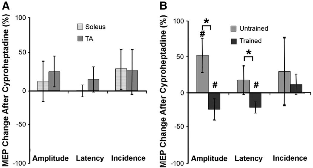
Spinal Wistar Hannover rats injected with olfactory ensheathing glia (OEG) have been shown to recover some bipedal stepping and climbing abilities. Given the intrinsic ability of the spinal cord to regain stepping with pharmacological agents or epidural stimulation after a complete mid-thoracic transection, we asked if functional recovery after OEG injections is due to changes in the caudal stump or facilitation of functional regeneration of axons across the transection site. OEG were injected rostral and caudal to the transection site immediately after transection. Robotically assisted step training in the presence of intrathecal injections of a 5-HT(2A) receptor agonist (quipazine) was used to facilitate recovery of stepping. Bipedal stepping as well as climbing abilities were tested over a 6-month period post-transection to determine any improvement in hindlimb functional due to OEG injections and/or step training. The ability for OEG to facilitate regeneration was analyzed electrophysiologically by transcranially stimulating the brainstem and recording motor evoked potentials (MEP) with chronically implanted intramuscular EMG electrodes in the soleus and tibalis anterior with and without intrathecal injections of noradrenergic, serotonergic, and glycinergic receptor antagonists. Analyses confirmed that along with improved stepping ability and increased use of the hindlimbs during climbing, only OEG rats showed recovery of MEP. In addition the MEP signals were eliminated after a re-transection of the spinal cord rostral to the original transection and were modified in the presence of receptor antagonists. These data indicate that improved hindlimb function after a complete transection was coupled with OEG-facilitated functional regeneration of axons. This article is part of a Special Issue entitled: Understanding olfactory ensheathing glia and their prospect for nervous system repair.


Figures

Fig. 1
Fig. 1
Stepping and climbing apparatus. A shows the robotically assisted step training method (Ziegler et al., 2010). If the ankle of the rat is at a position within the open circle (marked by the “X”) the robotic arms are passive and the leg is able to move freely in any direction within that area (open arrows). When the ankle leaves the circle the robotic arms guide it back into the open circle. The robotic arms also provide a small force guiding the ankle to the next point (closed circle) in the programmed trajectory. The closed arrows show forces that adjust the ankle position to this area which are proportional to the distance from the desired position. B shows the apparatus used for the climb test (Ramón-Cueto et al., 2000). A platform (50 cm × 30 cm) with a slippery surface was set up 70 cm from the floor and a wire grid (2.5 cm grid holes) was set at three different inclines (45°, 60°, and 90°) during testing.
Fig. 2
Fig. 2
Improved stepping through training and 5-HT agonists for Media rats tested (20 s) after 4 months of training post-transection. Panels A–D show examples of different kinematics aspects of stepping for Media rats under three conditions: after injections of quipazine alone (+Q), left column; after step training (+T) plus injection of quipazine, center column; and after step training plus quipazine plus 8-OH-DPAT (+8), right column. A shows a stick figure diagram of a representative step under each condition, from markers at the iliac crest, hip, knee, ankle, foot, and toe. The two examples from the trained groups are from the same trained rat. B shows the average step trajectory, taken from all completed steps in one session, for the rats shown in A. C shows the EMG activity in the soleus and TA muscles from a single plantar step and D shows the average EMG of 10 steps from the rats shown in A–C. E shows the average number of plantar placements taken during 20 s of stepping by each of the same three groups as above plus a Media group receiving no interventions. F shows the average PCA analysis (see Methods) for the same groups as in E. G shows the mean height and length of the step trajectory. H shows the average percent time co-activation of the soleus and TA muscles. Bars in E–H are mean ± SEM for 10 rats/group, except +Q, +T, and +8 which is a group of 7 rats. *, significant difference between groups at p<0.05.
Fig. 3
Fig. 3
Step kinematics of Media and OEG rats taken 6 months post-transection, unless otherwise noted. A–D show a stick figure diagram of a typical step from a representative rat from each group. Underneath each stick figure is the averaged raw EMG activity of the soleus and TA during stepping for each rat for one testing session, except for the Media trained as no rat from this group stepped in months four through six (N/A). E shows the average number of plantar placements taken by the Media untrained (M-U), Media trained (M-T), OEG untrained (O-U), and OEG trained (O-T) groups over the 6-month experimental period. F shows the average PCA analysis for each group of rats over all testing sessions. G shows the average height and length for all plantar placements taken by each group over all testing sessions. H shows the total number of OEG and Media rats that took plantar placement steps each month. I shows the average number of plantar placements taken by OEG and Media rats during each month. J shows the percent time of co-activation of the soleus and TA for the averaged step EMG data shown in A, B, and D. Bars in E–G and J are mean ± SEM for 10 rats/group. Bars in I are ± SEM for 15–20 rats/group. *, significant difference between groups at p<0.05.
Fig. 4
Fig. 4
Stepping ability of Media and OEG rats with quipazine administration. A shows the PCA analysis and B the number of plantar placement steps for each group for 20 s of stepping after quipazine administration. Bars are mean ± SEM for 10 rats/group. Group abbreviations, same as in Fig. 3. *, significant difference between groups at p<0.05.
Fig. 5
Fig. 5
Hindlimb usage during climbing for Media untrained and OEG untrained rats A shows the average number of times that each of the OEG or Media rats pushed off with their hindlimbs during the climbing task. B shows the mean (±SEM) EMG amplitude of the TA and soleus muscles in the OEG and Media groups for the pushoff and at all other times (spontaneous) during the climbing task. All tests were done on 6 rats per group. *, significant difference between OEG and Media groups at p<0.05. †, significant difference within the OEG group at p<0.05.
Fig. 6
Fig. 6
Examples of MEP at different time points during the experiment. A shows a MEP response to brainstem stimulation for an intact rat. The recording saturated at 10 mV. Therefore a flat portion of the signal appears if the values are higher than 10 mV. B is a MEP response for an OEG trained rat 1 month post-transection. C is the recovered MEP in the OEG rat in B 6 months post-transection. D is a MEP response 6 months post-transection in an OEG untrained rat. E shows the lack of any MEP response 6 months post-transection in a Media rat. F shows the lack on a MEP response from the same rat in graphs B and C after re-transection of the spinal cord at a level rostral to the initial transection. The scale bar in C is the same for B–F.
Fig. 7
Fig. 7
MEP amplitudes relative to hindlimb performance in trained and untrained OEG rats 6 months post-transection. A and B show the relationship between the number of times the hindlimbs were used (pushing off) and the normalized MEP amplitude ((MEP Amplitude (mV)/Max Stepping EMG (mV)) × 100)) in the soleus and TA muscles of untrained OEG rats, respectively. Each dot represents the number of pushoffs for a single leg (2 dots per rat). C and D show the relationship between the number of plantar placement steps taken by each trained and untrained OEG rat and the normalized MEP amplitude of the soleus and TA muscles, respectively. E–G show the differences in the normalized amplitude (E), latency (F), and incidence (G) of the MEP response between OEG trained and untrained rats. The correlation plots are based on data from 6 OEG untrained rats (A and B) or 8 OEG trained and 5 OEG untrained rats (C–D). Bar graphs in (E–G) are means ± SEM for 8 OEG trained and 5 OEG untrained rats.*, significant difference between groups at p<0.05.
Fig. 8
Fig. 8
MEP modulation due to yohimbine, a NA2 receptor antagonist. A and B show the MEP response of all OEG rats after intrathecal injection of yohimbine. A shows the percent change in MEP amplitude, latency, and incidence for each muscle post- compared to pre-injection. B shows the percent change in MEP amplitude, latency, and incidence post-compared to pre-injection in both muscles of OEG trained and untrained rats. Bars are mean ± SEM for 7 rats. #, significant difference between pre- and post-yohimbine injection at p<0.05.
Fig. 9
Fig. 9
MEP modulation due to cyproheptadine, a 5-HT2 receptor antagonist. A and B show the MEP response of all OEG rats after intrathecal injection of cyproheptadine. A shows the percent change in MEP amplitude, latency, and incidence for each muscle post- compared to pre-injection. B shows the percent change in MEP amplitude, latency, and incidence post- compared to pre-injection in both muscles of OEG trained and untrained rats. Bars are mean ± SEM for 5 rats. *, and # significant difference between groups and between pre-and post-cyproheptadine injection, respectively, at p<0.05.
Fig. 10
Fig. 10
MEP modulation due to strychnine, a glycinergic receptor antagonist. A and B show the MEP response of all OEG rats after intrathecal injection of strychnine. A shows the percent change in MEP amplitude, latency, and incidence for each muscle post- compared to pre-injection. B shows the percent change in MEP amplitude, latency, and incidence post- compared to pre-injection in both muscles of OEG trained and untrained rats. Bars are mean ± SEM for 7 rats. *, and #, significant difference between groups and between pre-and post-strychnine injection, respectively, at p<0.05.
Fig. 11
Fig. 11
Graphic summary of the effects of each receptor antagonist on the amplitude of the MEP response in untrained (UTr) and trained (Tr) OEG rats as shown in Figs. 8–10.

Similar articles

Cited by

References

    1. Cai LL, Fong AJ, Otoshi CK, Liang Y, Burdick JW, Roy RR, Edgerton VR. Implications of assist-as-needed robotic step training after a complete spinal cord injury on intrinsic strategies of motor learning. J. Neurosci. 2006;26:10564–10568. - PMC - PubMed
    1. Boulenguez P, Liabeuf S, Bos R, Bras H, Jean-Xavier C, Brocard C, Stil A, Darbon P, Cattaert D, Delpire E, Marsala M, Vinay L. Down-regulation of the potassium-chloride cotransporter KCC2 contributes to spasticity after spinal cord injury. Nat. Med. 2010;16:302–307. - PubMed
    1. de Leon RD, Acosta CN. Effect of robotic-assisted treadmill training and chronic quipazine treatment on hindlimb stepping in spinally transected rats. J. Neurotrama. 2006;23:1147–1163. - PubMed
    1. de Leon RD, Tamaki H, Hodgson JA, Roy RR, Edgerton VR. Hindlimb locomotor and postural training modulates glycinergic inhibition in the spinal cord of the adult spinal cat. J. Neurophysiol. 1999;82:359–369. - PubMed
    1. Edgerton VR, Roy RR. Spasticity: a switch from inhibition to excitation. Nat. Med. 2010;16:270–271. - PubMed

Publication types