. 2011 Apr 5;84(6):376-80.
doi: 10.1016/j.brainresbull.2011.01.006. Epub 2011 Jan 25.

TPH2 in the ventral tegmental area of the male rat brain

Affiliations

TPH2 in the ventral tegmental area of the male rat brain

Nurgul Carkaci-Salli et al. Brain Res Bull. .

Abstract

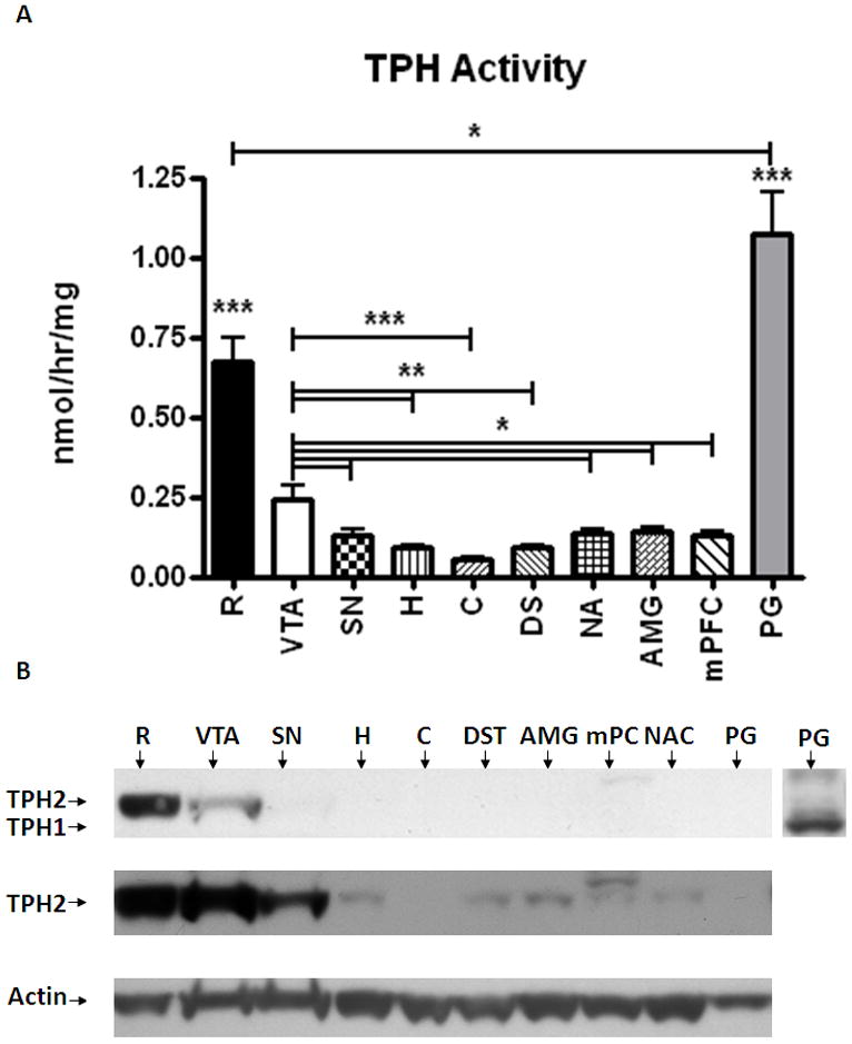
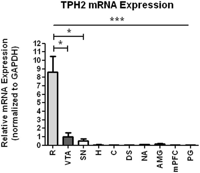
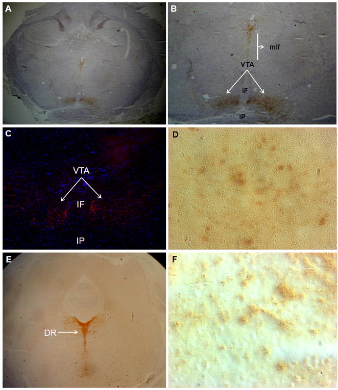
This study surveyed the distribution of tryptophan hydroxylase 2 (TPH2) mRNA, protein, and enzymatic activity throughout the male Sprague-Dawley rat brain. TPH2 is the genetic isoform of TPH that catalyzes the rate-limiting step in serotonin biosynthesis within the central nervous system. Although cell bodies of serotonergic neurons are located mainly in the raphe, serotonin-containing axons innervate many regions of the brain. In the present study, we assessed the levels of mRNA, protein expression, and enzyme activity of TPH2 in the rat raphe, ventral tegmental area (VTA), substantia nigra, hippocampus, cerebellum, dorsal striatum, nucleus accumbens, amygdala, and medial prefrontal cortex to more fully understand the distribution of this enzyme throughout the central nervous system. The pineal gland was used as a control tissue that expresses TPH1 (the peripheral enzyme), but not TPH2. As expected, the raphe showed the highest brain TPH2 activity and protein expression. In the contrast to other reports, however, the VTA followed the raphe as the region with the second-highest amount of TPH2 activity, mRNA and protein expression. There were significantly lower TPH activities and levels of TPH2 protein in the other regions. In addition, TPH2 immunocytochemistry demonstrated the presence of TPH-positive cell bodies within the VTA. The results of this study indicate that TPH2 and serotonergic signaling may play an important role in the mesolimbic/mesocortical reward pathway.


Figures

Figure 1
Figure 1
A- Relative TPH2 activity was measured with a radio-enzymatic assay in brain homogenates from raphe (R), ventral tegmental area (VTA), substantia nigra (SN), hippocampus (H), cerebellum (C), dorsal striatum (DST), amygdala (AMG), medial prefrontal cortex (mPFC), and nucleus accumbens (NAc). The pineal gland (PG) served as negative control, with no TPH2. Activity levels were normalized to the total protein levels in the homogenates. Data expressed as mean activity/total protein ±SEM. GraphPad Prism5 was used for one-way ANOVA with a Newman-Keuls post-hoc test. Statistical significance * p<0.05, ** p<0.01, *** p<0.001, n=8. B- Determination of TPH2 expression levels in different brain regions used for the TPH activity assay. An aliquot of total protein (20μg) was loaded into each lane. The inset at the right represents the same pineal gland portion of the blot probed with an antibody that detects both TPH1 (peripheral) and TPH2 (central) isoforms of the enzyme. Top panel; TPH2-specific rabbit anti-TPH2 antibody (dilution: 1:5000). Middle panel; longer exposure of the blot presented above. Lower panel; β-actin bands were used as loading control to normalize TPH2 expression to protein levels.
Figure 2
Figure 2
Relative TPH2 protein levels in raphe and VTA tissue homogenates were quantified by measuring the optical density of the TPH2 bands detected by western blot using TPH2-specific rabbit anti-TPH2 antibody (dilution: 1:5000) and normalized to the actin intensity. Results were analyzed by using GraphPad Prism5 one-way ANOVA with Newman-Keuls post-hoc test. The error bars represent standard error of the mean. Statistical significance * p<0.05, ** p<0.01, *** p<0.001, n=8.
Figure 3
Figure 3
TPH2 mRNA expression in brain tissue from raphe (R), ventral tegmental area (VTA), substantia nigra (SN), hippocampus (H), cerebellum (C), dorsal striatum (DST), amygdala (AMG), medial prefrontal cortex (mPFC), nucleus accumbens (NAc), and pineal gland (PG). TPH2 mRNA expression levels were normalized to the expression levels of GAPDH in the samples. Results were analyzed using GraphPad Prism5 one-way ANOVA with Newman-Keuls post-hoc test. The error bars represent standard error of the mean. Statistical significance * p<0.05, ** p<0.01, *** p<0.001, n=2-4.
Figure 4
Figure 4
Immunocytochemistry assessment of TPH2 expression in the VTA region using rabbit anti-TPH2 specific antibody and HRP- (A,B,D,E and F) and immunofluorescent-conjugated secondary antibodies (C). A- TPH2 positive neuronal bodies are located within the VTA region (areas indicated by arrows), but not the surrounding areas such as the interfascicular nucleus (IF) and interpeduncular (IP) nucleus. In addition to the VTA, TPH2-positive neurons were found in the medial longitudinal fasciculus (MLF). B-Enlarged view of the VTA region. C- Immunofluorescent detection of TPH2 positive neurons (arrows) in the VTA region from an independent section using the same antibody. D- Enlarged view of the VTA region displays TPH2–positive neuronal bodies in the VTA. E- TPH2 positive neuronal bodies are located within the dorsal raphe (DR) region (areas indicated by arrows). F- Enlarged view of DR region displays TPH2–positive neuronal bodies in the DR.

References

    1. Baker KG, Halliday GM, Hornung JP, Geffen LB, Cotton RG, Tork I. Distribution, morphology and number of monoamine-synthesizing and substance P-containing neurons in the human dorsal raphe nucleus. Neuroscience. 1991;42:757–775. - PubMed
    1. Bendotti C, Servadio A, Samanin R. Distribution of GAP-43 mRNA in the brain stem of adult rats as evidenced by in situ hybridization: localization within monoaminergic neurons. J Neurosci. 1991;11:600–607. - PMC - PubMed
    1. Bowyer JF, Pogge AR, Delongchamp RR, O’Callaghan JP, Patel KM, Vrana KE, Freeman WM. A threshold neurotoxic amphetamine exposure inhibits parietal cortex expression of synaptic plasticity-related genes. Neuroscience. 2007;144:66–76. - PMC - PubMed
    1. Deguchi T, Barchas J. Effect of p-chlorophenylalanine on hydroxylation of tryptophan in pineal and brain of rats. Mol Pharmacol. 1972;8:770–779. - PubMed
    1. Deguchi T, Barchas J. Regional distribution and developmental change of tryptophan hydroxylase activity in rat brain. J Neurochem. 1972;19:927–929. - PubMed

Publication types