

Polyethylenimine-conjugated gold nanoparticles: Gene transfer potential and low toxicity in the cornea
- PMID: 21272669
- PMCID: PMC3094737
- DOI: 10.1016/j.nano.2011.01.006
Polyethylenimine-conjugated gold nanoparticles: Gene transfer potential and low toxicity in the cornea
Abstract
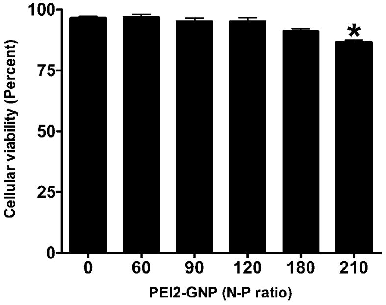
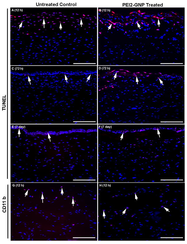
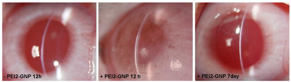
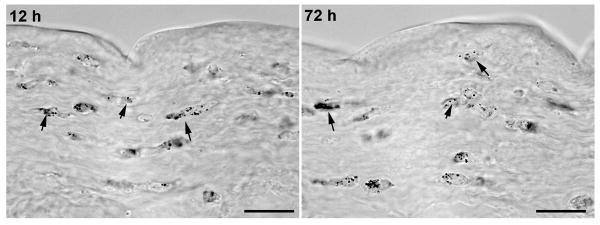
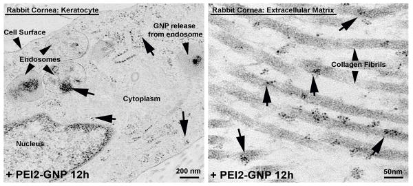
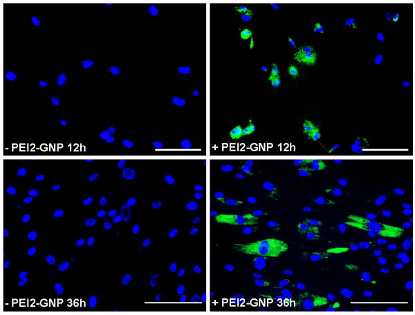
This study examined the gene transfer efficiency and toxicity of 2-kDa polyethylenimine conjugated to gold nanoparticles (PEI2-GNPs) in the human cornea in vitro and rabbit cornea in vivo. PEI2-GNPs with nitrogen-to-phosphorus ratios of up to 180 exhibited significant transgene delivery in the human cornea without altering the viability or phenotype of these cells. Similarly, PEI2-GNPs applied to corneal tissues collected after 12 hours, 72 hours, or 7 days exhibited appreciable gold uptake throughout the rabbit stroma with gradual clearance of GNPs over time. Transmission electron microscopy detected GNPs in the keratocytes and the extracellular matrix of the rabbit corneas. Additionally, slit-lamp biomicroscopy in live animals even 7 days after topical PEI2-GNP application to the cornea detected no inflammation, redness, or edema in rabbit eyes in vivo, with only moderate cell death and immune reactions. These results suggest that PEI2-GNPs are safe for the cornea and can potentially be useful for corneal gene therapy in vivo.
From the clinical editor: This study examined the gene transfer efficiency and toxicity of 2-kDa polyethylenimine conjugated to gold nanoparticles in the human cornea in vitro and rabbit cornea in vivo. The results suggest that PEI2-GNPs are safe for the cornea and can potentially be useful for corneal gene therapy in vivo.
Published by Elsevier Inc.
Conflict of interest statement
Conflict of Interest Statement: None of the authors have any conflict of interest.
Figures
References
-
- Mohan RR, Sharma A, Netto MV, Sinha S, Wilson SE. Gene therapy in the cornea. Prog Retin Eye Res. 2005;24:537–59. - PubMed
-
- Sharma A, Ghosh A, Siddapa C, Mohan RR. Ocular Surface: Gene Therapy. In: Besharse J, Dana R, Dartt DA, editors. Encyclopedia of the Eye. Elsevier; 2010. pp. 185–194.
Publication types
MeSH terms
Substances
Grants and funding
LinkOut - more resources
Full Text Sources
Other Literature Sources
