. 2011 Apr;105(4):1506-15.
doi: 10.1152/jn.00950.2010. Epub 2011 Jan 27.

Cc2d1a, a C2 domain containing protein linked to nonsyndromic mental retardation, controls functional maturation of central synapses

Affiliations

Cc2d1a, a C2 domain containing protein linked to nonsyndromic mental retardation, controls functional maturation of central synapses

Meng Zhao et al. J Neurophysiol. 2011 Apr.

Abstract

Cc2d1a is an evolutionarily conserved protein composed of NH(2)-terminal Drosophila melanogaster 14 domain (DM14) domains and a COOH-terminal C2 domain. Human patients with homozygotic mutation in the gene suffer from nonsyndromic mental retardation, implying that Cc2d1a functions in the central nervous system. To examine the physiological role of the Cc2d1a, we generated and analyzed Cc2d1a knockout (KO) mice. Cc2d1a KO mice die soon after birth, apparently because of their inability to breathe. Histological analysis of Cc2d1a KO animals did not identify any structural defects in the peripheral respiratory apparatus. However, functional analysis of synapses formed between Cc2d1a-deficient cortical neurons revealed a robust increase in the pace of maturation of evoked synaptic responses as well as synaptic vesicle trafficking. This synaptic anomaly was rescued by reintroducing full-length Cc2d1a but not C2-domain-deletion mutant, underscoring the functional importance of C2 domain. Our data suggest that Cc2d1a is required for mouse survival and performs essential function in controlling functional maturation of synapses.


Figures
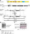
Fig. 1.
Fig. 1.
Targeting the Cc2d1a locus. A: strategy to generate the Cc2d1a-deficient allele. The domain structure of the mouse (m) Cc2d1a protein, first 3 ATG sites, corresponding exonal structure, targeting vector, and targeted allele are shown. LacZ cassette and loxP-Neo-loxP replace exons 1–10. Positions of the probe used for Southern analysis are shown. B: genomic DNA from mice of the indicated genotypes was analyzed by Southern blot. C: protein extracted from E14.5 myocyte enhancer factor cells was analyzed by immunoblotting using CC2D1A-specific antibody. Genomic DNA from embryonic day 14.5 (E14.5) embryos was analyzed by genotyping PCR. D: number of E18.5 embryos from the breeding of Cc2d1a+/−. WT, wild-type; KO, knockout; IB: immunoblot; Het, heterozygous mice.
Fig. 2.
Fig. 2.
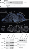
Expression of Cc2d1a is enriched in the brain. A: protein extracted from different tissues of E18.5 embryos were examined by Western blot using CC2D1A antibody. B: detection of Cc2d1a transcripts by in situ hybridization. Inset: sagittal section of a KO embryo. In the center is a sagittal section of a WT embryo. At the bottom are coronal sections of a WT embryo. C: subcellular fractionation of adult forebrains using Percoll step gradient. As described in methods, brain homogenate was centrifuged at 1,000 g to obtain low-speed supernatant (Sup) (S1) or 100,000 g to obtain cytosol. Low-speed supernatant was centrifuged at 14,500 g, and the pellet provided crude synaptosomes. Synaptosome fraction banded at the interface of 12%/20% Percoll. The same amount of protein from each fraction was used for Western blotting. Protein extract from embryonic brain tissues of WT and KO mice were used to verify the specificity of the Cc2d1a antibody. MW, molecular weight; NMDAR-1, N-methyl-d-aspartate receptor-1; Syt-1, Synaptotagmin-1; Syp-1, Synaptophysin-1; Stx-1, Syntaxin-1.
Fig. 3.
Fig. 3.
Anatomy of Cc2d1a KO mice is normal. A: top: sagittal sections of WT and Cc2d1a KO embryos at E18.5. Bottom, left: transverse sections through the heart and lung. Bottom, right: coronal sections through the brain. B: whole-mount embryonic diaphragm muscles (E18.5) were immunostained with anti-Syntaxin antibody. C: confocal images of single endplates from whole-mount diaphragm muscle at E18.5, double-labeled with Syt-2 antibody (green) and Texas-red conjugated α-bungarotoxin (red).
Fig. 4.
Fig. 4.
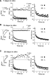
Evoked GABAergic responses in Cc2d1a KO neurons. A: representative traces (inset and left) and average synaptic responses (right) from 7 days in vitro (DIV) cortical neurons. Evoked inhibitory postsynaptic currents (IPSCs) were recorded in response to maximal field stimulation applied for 5 s at 10 Hz. The mean amplitudes of GABAergic responses (IPSCs) under the inhibition of glutamatergic responses were measured at each stimulation (Control, n = 14; KO, n = 13). Representative traces on the left depict the first responses from control or Cc2d1a KO neurons. P values indicating the statistical significance of the difference between control and Cc2d1a KO traces are plotted above the average values. When IPSCs were normalized with respect to the first response (IPSCmax) the resulting traces did not reveal a kinetic difference between the control and KO conditions. B: cortical neurons at 14 DIV were examined as in A (Control, n = 20; KO n = 22). The difference between the average traces is significant (P < 0.05) throughout the stimulation period. Inset: representative traces (scale bar 1 s, 0.2 nA). C: cortical neurons at 21 DIV were examined as in A (Control, n = 6; KO, n = 7). Inset: representative traces (scale bar 1 s, 1 nA). Note that control and KO groups do not show a clear difference at this stage. In all cases, the normalized IPSC values (shown on the right) reveal no differences in the decay kinetics between the groups at 7, 14 or 21 DIV.
Fig. 5.
Fig. 5.
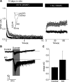
Evoked excitatory neurotransmission in Cc2d1a KO neurons. A: top: average of excitatory postsynaptic currents (EPSCs) evoked at 14 DIV cultures of control or Cc2d1a KO neurons. AP, action potential. Bottom: all the 20-Hz stimulation peaks were significantly different between the 2 groups. Inset: representative traces for the initial peaks of control and Cc2d1a KO groups. At the end of the 20-Hz stimulation paradigm, switching to 1-Hz stimulation enabled rapid recovery of both control and Cc2d1a KO synaptic response to their initial values (n = 11 for control and n = 10 for KO cells). B and C: representative traces (B) and mean charge movement values integrated during the first 10 s of 20-s hypertonic stimulation (with the addition of 0.5 M sucrose) of excitatory synapses (C) in control and Cc2d1a KO neurons. These experiments did not reveal a significant difference between the 2 groups (n = 6 for both, P > 0.05).
Fig. 6.
Fig. 6.
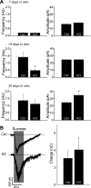
Analysis of spontaneous release events and hypertonic sucrose-triggered responses of GABAergic synapses in Cc2d1a-deficient neurons. A: cultured cortical neurons at 7 (top), 14 (middle), and 21 (bottom) DIV were analyzed in voltage-clamp mode in the presence of 1 μM tetrodotoxin. Comparison of miniature IPSC (mIPSC) frequencies (left) and amplitudes (right) for GABAergic events in control and Cc2d1a-deficient neurons (7 DIV: Control, n = 8 neurons, KO, n = 10; 14 DIV: Control, n = 16, KO, n = 11; 21 DIV: Control, n = 7, KO, n = 5) *Significant differences between control and KO cells. B: inhibitory responses evoked by hypertonic sucrose (+500 mOsm) application in control and Cc2d1a-deficient synapses (left). Sample recordings of inhibitory synaptic responses to a local application of +500 mM sucrose in Tyrode solution in the presence of 1 mM tetrodotoxin (right). The average values of total release induced by hypertonic sucrose application did not reveal a difference between control and KO conditions (Control, n = 3 neurons; KO, n = 4). The total charge transfer triggered by hypertonic sucrose application was determined by integrating the area over the first 10 s of 20-s stimulation.
Fig. 7.
Fig. 7.
Analysis of synaptic vesicle recycling using Synaptophysin-pHluorin. Cultured cortical neurons were infected with lentiviruses expressing synaptophysin-pHluorin at 4 DIV and analyzed at 14 DIV. A: left: representative experiment. Right: averaged traces (Control, n = 12 experiments; KO, n = 8). Electrical stimulation was applied at 20 Hz during 20 s. St, stimulation. Fluorescence changes were normalized to the peak value. The magnitude of the fluorescence change during stimulation represents the amount of synaptophysin-pHluorin accumulated on the synaptic plasma membrane. Fluorescence decay after stimulation represents endocytic retrieval of synaptophysin-pHluorin and reacidification of vesicles. ΔF = F0 (resting fluorescence)− Fmax (peak fluorescence). The average values are statistically different (P < 0.05) after the peak is reached, denoting a faster endocytosis in the KO neurons. B: left: fluorescence changes over baseline (F0) during stimulation or NH4Cl (50 mM) treatment do not show a statistically significant difference between control and KO synapses. AU, arbitrary units; Control, n = 5 experiments; KO, n = 4. Right: relative changes in fluorescence during stimulation with respect to the change after NH4Cl treatment is not different between the 2 groups. These data suggest that the number of vesicles that contain synaptophysin-pHluorin as well as the recycling fraction are not different between control and KO synapses despite a clear difference in the rate of endocytosis.
Fig. 8.
Fig. 8.
Cc2d1a partially rescues IPSCs in Cc2d1a-deficient neurons. A: cortical cultures were infected at 4 DIV with lentiviruses expressing either full-length (FL) or COOH-terminal-deletion mutant of Cc2d1a. The expression of exogenous proteins was examined by immunoblotting using CC2D1A-specific antibody. B: IPSC was examined at 14 DIV as described in Fig. 4 (Control, n = 17 neurons; KO, n = 15; KO-FL, n = 14; KO-ΔC, n = 8). We used ANOVA 1-way and Bonferroni or Fisher test to compare 4 groups of neurons. Expression of FL Cc2d1a in KO neurons significantly rescued IPSCs in response to the first and second action potential stimulation in the train compared with control neurons. The difference in evoked responses was not significant after the third action potential and beyond indicating partial rescue. However, ΔC-Cc2d1a construct did not rescue IPSCs throughout the stimulation train under all experimental conditions. C: graph depicts normalized IPSC amplitudes under all conditions. Depression kinetics did not reveal a significant difference.

References

    1. Agrawal N, Kango M, Mishra A, Sinha P. Neoplastic transformation, and aberrant cell-cell interactions in genetic mosaics of lethal(2)giant larvae (lgl), a tumor suppressor gene of Drosophila. Dev Biol 172: 218–229, 1995 - PubMed
    1. Akhtar MW, Raingo J, Nelson ED, Montgomery RL, Olson EN, Kavalali ET, Monteggia LM. Histone deacetylases 1, and 2 form a developmental switch that controls excitatory synapse maturation and function. J Neurosci 29: 8288–8297, 2009 - PMC - PubMed
    1. Atasoy D, Schoch S, Ho A, Nadasy KA, Liu X, Zhang W, Mukherjee K, Nosyreva ED, Fernandez-Chacon R, Missler M, Kavalali ET, Sudhof TC. Deletion of CASK in mice is lethal, and impairs synaptic function. Proc Natl Acad Sci USA 104: 2525–2530, 2007 - PMC - PubMed
    1. Barbosa AC, Kim MS, Ertunc M, Adachi M, Nelson ED, McAnally J, Richardson JA, Kavalali ET, Monteggia LM, Bassel-Duby R, Olson EN. MEF2C, a transcription factor that facilitates learning, and memory by negative regulation of synapse numbers and function. Proc Natl Acad Sci USA 105: 9391–9396, 2008 - PMC - PubMed
    1. Basel-Vanagaite L, Attia R, Yahav M, Ferland RJ, Anteki L, Walsh CA, Olender T, Straussberg R, Magal N, Taub E, Drasinover V, Alkelai A, Bercovich D, Rechavi G, Simon AJ, Shohat M. The CC2D1A, a member of a new gene family with C2 domains, is involved in autosomal recessive non-syndromic mental retardation. J Med Genet 43: 203–210, 2006 - PMC - PubMed

Publication types

MeSH terms