. 2011 Jan 26;31(4):1292-301.
doi: 10.1523/JNEUROSCI.4611-10.2011.

PreBotzinger complex neurokinin-1 receptor-expressing neurons mediate opioid-induced respiratory depression

Affiliations

PreBotzinger complex neurokinin-1 receptor-expressing neurons mediate opioid-induced respiratory depression

Gaspard Montandon et al. J Neurosci. .

Abstract

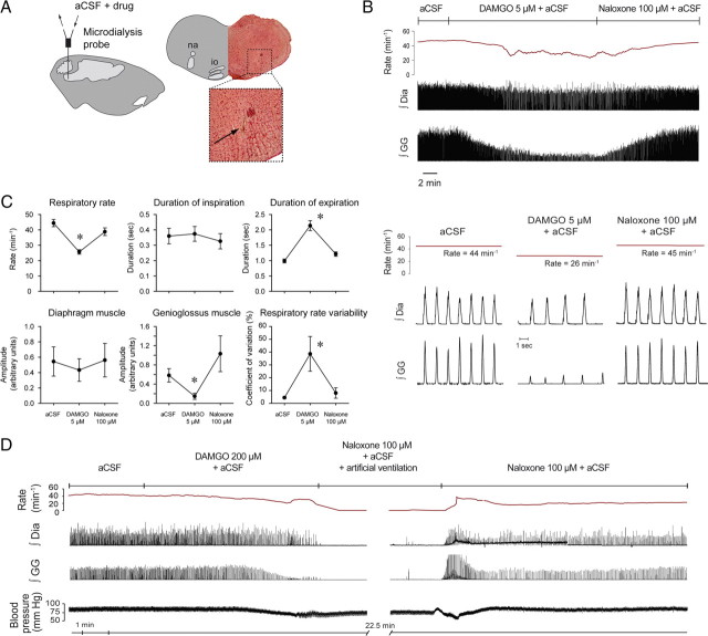
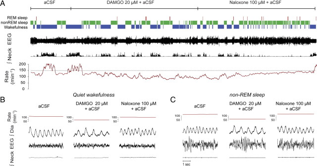
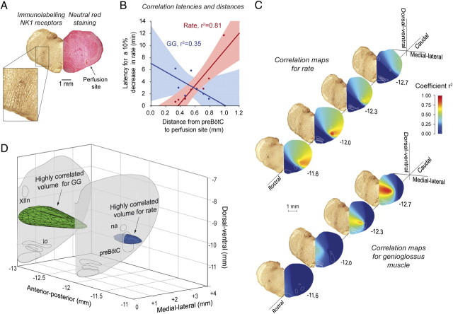
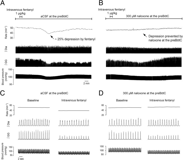
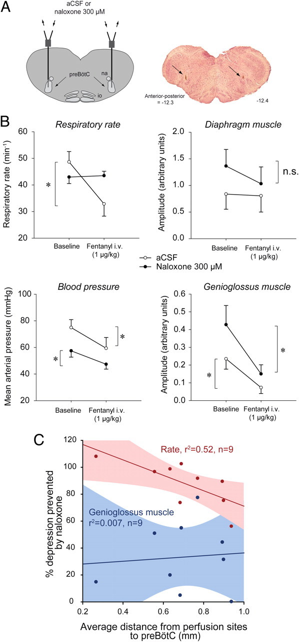
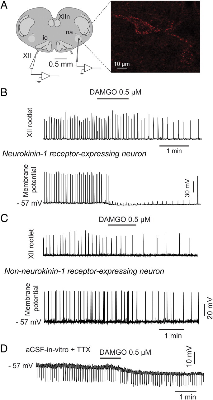
The analgesic properties of the opium poppy Papever somniferum were first mentioned by Hippocrates around 400 BC, and opioid analgesics remain the mainstay of pain management today. These drugs can cause the serious side-effect of respiratory depression that can be lethal with overdose, however the critical brain sites and neurochemical identity of the neurons mediating this depression are unknown. By locally manipulating neurotransmission in the adult rat, we identify the critical site of the medulla, the preBötzinger complex, that mediates opioid-induced respiratory depression in vivo. Here we show that opioids at the preBötzinger complex cause respiratory depression or fatal apnea, with anesthesia and deep-sleep being particularly vulnerable states for opioid-induced respiratory depression. Importantly, we establish that the preBötzinger complex is fully responsible for respiratory rate suppression following systemic administration of opioid analgesics. The site in the medulla most sensitive to opioids corresponds to a region expressing neurokinin-1 receptors, and we show in rhythmically active brainstem section in vitro that neurokinin-1 receptor-expressing preBötzinger complex neurons are selectively inhibited by opioids. In summary, neurokinin-1 receptor-expressing preBötzinger complex neurons constitute the critical site mediating opioid-induced respiratory rate depression, and the key therapeutic target for its prevention or reversal.


Figures

Figure 1.
Figure 1.
Opioid receptor mechanisms at the preBötC modulate breathing in vivo. A, Schema and histological section showing unilateral location of the tip of the microdialysis probe (arrow) in the medulla. B, Example showing that addition of 5 μm DAMGO to the aCSF perfusing the preBötC caused respiratory depression, effects that were reversed with naloxone. C, Group mean data (n = 9) for the effect of DAMGO (5 μm), and then the subsequent reversal of effects with naloxone (100 μm), on respiratory rate, duration of inspiration, duration of expiration, the amplitude of diaphragm (Dia) muscle activation, the amplitude of genioglossus (GG) muscle activation, and respiratory rate variability. Error bars are SEM. D, DAMGO (200 μm) caused complete respiratory arrest in the anesthetized adult rat. The animal was kept alive by artificial ventilation, and respiratory activity recovered after 30 min of perfusion with naloxone (100 μm). *p < 0.05, significant differences compared with aCSF controls. na, Nucleus ambiguus; io, inferior olive.
Figure 2.
Figure 2.
In vivo modulation of breathing by the μ-opioid agonist DAMGO at the preBötC across sleep–wake states. A, Recordings of a whole experimental protocol with traces showing the prevailing sleep–wake states, electroencephalogram (EEG) activity, integrated neck muscle activity, mean respiratory rate (derived from successive 10 s time-bins), instantaneous (breath-by-breath) rate, and integrated diaphragm (Dia) muscle activity. The top line represents the time course for the continuous perfusion of the different drugs into the preBötC, with the dashed lines showing the 30 min periods used to calculate the mean data used in Figure 3. Note the persistent suppression of respiratory rate during perfusion of DAMGO, which is especially apparent during non-REM sleep, and the subsequent reversal of this rate depression by naloxone. B, C, DAMGO (20 μm) at the preBötC caused respiratory slowing in wakefulness (B) and non-REM sleep (C), with this effect reversed by naloxone.
Figure 3.
Figure 3.
Bilateral microdialysis perfusion of the μ-opioid receptor agonist DAMGO into the preBötC of freely behaving adult rats. A, Bilateral sites of perfusion (tips of the microdialysis probes indicated by the arrows) identified from a section of medulla at 12.3 mm posterior to bregma. B, Group mean data for the effect of DAMGO (20 μm) at the preBötC on respiratory rate and the amplitude of diaphragm muscle activation across states in conscious freely behaving rats (n = 3). Error bars are SEM. The reversal of effects by bilateral application of the μ-opioid receptor antagonist naloxone (100 μm) is also shown. Note the significant depression of respiratory rate but not diaphragm muscle amplitude by DAMGO at the preBötC. C, Significant relationship between the degree of opioid-induced respiratory rate depression and the depth of sleep as indexed by the ratio of high (β2, 20–30 Hz) to low (δ1, 0.5–2 Hz) EEG frequencies. *p < 0.05, values significantly different from aCSF.
Figure 4.
Figure 4.
Identification of the brainstem sites affected by opioids. A, Site of microdialysis perfusion located in the preBötC region as identified by neurokinin-1 receptor (NK1R) expression (n = 3) and histology (n = 8). B, Correlations between distances from the preBötC (−12.3 mm posterior, 2.5 mm lateral, and 10.2 mm ventral to bregma) to each perfusion site, and the corresponding latencies to a 10% decrease in respiratory rate (shown in red) or genioglossus (GG) muscle amplitude (blue). Note that the correlation between the perfusion sites and the corresponding latencies for respiratory rate depression is high and statistically significant (red, r2 = 0.81, p = 0.002), but for genioglossus activity the correlation is weak and not significant (blue, r2 = 0.35, p = 0.164). See supplemental Table 1, available at www.jneurosci.org as supplemental material for additional information. C, Such correlations were performed for all regions on the histological sections using a 50 μm square grid, and the magnitude of these correlations are color coded. These correlation maps reveal “hot spots” for the region most strongly associated with opioid-induced respiratory rate suppression, with this region corresponding to the preBötC. A similar approach for a 10% suppression of GG activity shows that a distinct region located dorsomedial and caudal to the preBötC was most strongly associated with opioid-induced suppression of tongue muscle activity (supplemental Fig. 1F,G, available at www.jneurosci.org as supplemental material). D, Three-dimensional reconstruction showing only those regions with high correlations (r2 > 0.8), with the regions associated with suppression of respiratory rate (blue) and GG activity (green) being nonoverlapping. XIIn, Hypoglossal motor nucleus; io, inferior olive; na, nucleus ambiguus.
Figure 5.
Figure 5.
The preBötC mediates opioid-induced respiratory rate suppression following systemic administration of fentanyl. A, With aCSF at the preBötC, intravenous application of fentanyl (1 μg/kg over 1 min) caused suppression of respiratory rate, genioglossus (GG) muscle activity and blood pressure. B, With naloxone at the preBötC, the suppression of respiratory rate following intravenous application of fentanyl (1 μg/kg over 1 min) was fully prevented, whereas the suppression of GG muscle activity and blood pressure still occurred. C, D, Tracings in a shorter time scale. Dia, diaphragm muscle amplitude.
Figure 6.
Figure 6.
Prevention of opioid-induced respiratory depression by naloxone at the preBötC in anesthetized rats (n = 4). A, Schema and histological section showing location of probes (arrows) for bilateral application of aCSF or naloxone into both preBötCs. B, Full prevention of fentanyl-induced suppression of respiratory rate by naloxone (300 μm, filled circles compared with aCSF (control, open circles). Diaphragm muscle amplitude was slightly, but not significantly, reduced by intravenous administration of fentanyl. Naloxone at the preBötC failed to prevent the suppression of blood pressure and genioglossus muscle activity produced by intravenous fentanyl. Data are means ± SEM. C, Significant correlation (r2 = 0.52, p = 0.028) between degree of prevention of respiratory rate and average distance from perfusion sites to preBötCs (−12.5 mm posterior, 2.5 mm either side of the midline, and −10.2 mm ventral to bregma). These coordinates correspond to a region of high NK1R expression. Genioglossus muscle suppression by opioids was not correlated with the average distance from perfusion sites to preBötCs since naloxone did not prevent the suppression. supplemental Figure 2 (available at www.jneurosci.org as supplemental material) shows that respiratory rate depression following intravenous opioids was not fully prevented even when one of the two naloxone perfusion sites was at the preBötC, i.e., both are required and both mediate the suppression following systemic opioid administration. * indicates when fentanyl significantly decreases activity (p < 0.05, post hoc Bonferroni t tests).
Figure 7.
Figure 7.
μ-Opioid receptor-induced inhibition of neurokinin-1 receptor-expressing (NK1R) inspiratory preBötC neurons in vitro. A, Whole-nerve recording from hypoglossal (XII) rootlet and whole-cell intracellular recording of NK1R-expressing preBötC neurons (red fluorescent labeling from internalization of tetramethylrhodamine-conjugated Substance P). B, Application of DAMGO (0.5 μm) to the bathing medium of the tissue slice decreased respiratory rate (as shown by decreased frequency of respiratory bursts in the XII nerve rootlet). Simultaneous current-clamp recording of a NK1R-expressing preBötC neuron showed membrane hyperpolarization, and clear suppression of inspiratory depolarization such that the threshold for action potential generation was not reached. C, Recordings of a non-NK1R-expressing preBötC neuron showing its lack of response to bath application of the μ-opioid receptor agonist DAMGO (0.5 μm). D, DAMGO-induced membrane hyperpolarization persisted in the presence of TTX (1 μm). na, Nucleus ambiguus; io, inferior olive, XIIn, hypoglossal motor nucleus; XII hypoglossal nerve rootlet.

Similar articles

Cited by

References

    1. Agrò F, Salvinelli F, Casale M, Gherardi S. Difficulty in airway management during sedation of patients affected by obstructive sleep apnea. Can J Anaesth. 2004;51:279. - PubMed
    1. Ballanyi K, Panaitescu B, Ruangkittisakul A. Indirect opioid actions on inspiratory pre-Botzinger complex neurons in newborn rat brainstem slices. Adv Exp Med Biol. 2010;669:75–79. - PubMed
    1. Bilkei-Gorzo A, Berner J, Zimmermann J, Wickström R, Racz I, Zimmer A. Increased morphine analgesia and reduced side effects in mice lacking the tac1 gene. Br J Pharmacol. 2010;160:1443–1452. - PMC - PubMed
    1. Desrosiers G. When opioid analgesia kills. Perspect Infirm. 2006;4:6–9. - PubMed
    1. Feldman JL, Del Negro CA. Looking for inspiration: new perspectives on respiratory rhythm. Nat Rev Neurosci. 2006;7:232–242. - PMC - PubMed

Publication types

MeSH terms